• Immunohistochemistry analysis of paraffin-embedded human brain tissue, using FAS ligand Antibody. The picture on the right is blocked with the synthesized peptide.
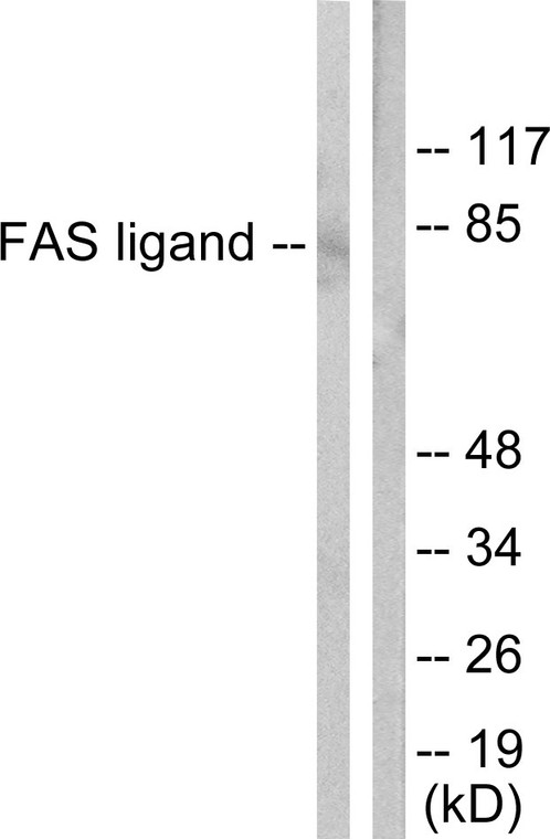
  • Western blot analysis of lysates from 293 cells, using FAS ligand Antibody. The lane on the right is blocked with the synthesized peptide.
  • Western blot analysis of various cells using FAS-L Polyclonal Antibody diluted at 1:1000
  • Western blot analysis of 293 cells using FAS-L Polyclonal Antibody diluted at 1:1000
  • Immunohistochemical analysis of paraffin-embedded Human-stomach-cancer tissue. 1, FAS-L Polyclonal Antibody was diluted at 1:200 (4°C, overnight). 2, Sodium citrate pH 6.0 was used for antibody retrieval (>98°C, 20min). 3, Secondary antibody was diluted at 1:200 (room tempeRature, 30min). Negative control was used by secondary antibody only.
  • Immunohistochemical analysis of paraffin-embedded Mouse-colon tissue. 1, FAS-L Polyclonal Antibody was diluted at 1:200 (4°C, overnight). 2, Sodium citrate pH 6.0 was used for antibody retrieval (>98°C, 20min). 3, Secondary antibody was diluted at 1:200 (room tempeRature, 30min). Negative control was used by secondary antibody only.
  • Immunohistochemical analysis of paraffin-embedded Human-colon tissue. 1, FAS-L Polyclonal Antibody was diluted at 1:200 (4°C, overnight). 2, Sodium citrate pH 6.0 was used for antibody retrieval (>98°C, 20min). 3, Secondary antibody was diluted at 1:200 (room tempeRature, 30min). Negative control was used by secondary antibody only.
  • Immunohistochemical analysis of paraffin-embedded Human-liver-cancer tissue. 1, FAS-L Polyclonal Antibody was diluted at 1:200 (4°C, overnight). 2, Sodium citrate pH 6.0 was used for antibody retrieval (>98°C, 20min). 3, Secondary antibody was diluted at 1:200 (room tempeRature, 30min). Negative control was used by secondary antibody only.
  • Immunofluorescence analysis of mouse-kidney tissue. 1, FAS-L Polyclonal Antibody (red) was diluted at 1:200 (4°C, overnight). 2, Cy3 labled Secondary antibody was diluted at 1:300 (room temperature, 50min).3, Picture B: DAPI (blue) 10min. Picture A:Target. Picture B: DAPI. Picture C: merge of A+B
  • Immunofluorescence analysis of mouse-kidney tissue. 1, FAS-L Polyclonal Antibody (red) was diluted at 1:200 (4°C, overnight). 2, Cy3 labled Secondary antibody was diluted at 1:300 (room temperature, 50min).3, Picture B: DAPI (blue) 10min. Picture A:Target. Picture B: DAPI. Picture C: merge of A+B
  • Immunofluorescence analysis of rat-kidney tissue. 1, FAS-L Polyclonal Antibody (red) was diluted at 1:200 (4°C, overnight). 2, Cy3 labled Secondary antibody was diluted at 1:300 (room temperature, 50min).3, Picture B: DAPI (blue) 10min. Picture A:Target. Picture B: DAPI. Picture C: merge of A+B
  • Immunofluorescence analysis of rat-kidney tissue. 1, FAS-L Polyclonal Antibody (red) was diluted at 1:200 (4°C, overnight). 2, Cy3 labled Secondary antibody was diluted at 1:300 (room temperature, 50min).3, Picture B: DAPI (blue) 10min. Picture A:Target. Picture B: DAPI. Picture C: merge of A+B
  • Immunofluorescence analysis of rat-lung tissue. 1, FAS-L Polyclonal Antibody (red) was diluted at 1:200 (4°C, overnight). 2, Cy3 labled Secondary antibody was diluted at 1:300 (room temperature, 50min).3, Picture B: DAPI (blue) 10min. Picture A:Target. Picture B: DAPI. Picture C: merge of A+B
  • Immunofluorescence analysis of rat-lung tissue. 1, FAS-L Polyclonal Antibody (red) was diluted at 1:200 (4°C, overnight). 2, Cy3 labled Secondary antibody was diluted at 1:300 (room temperature, 50min).3, Picture B: DAPI (blue) 10min. Picture A:Target. Picture B: DAPI. Picture C: merge of A+B
  • Western blot analysis of lysates from 1) HepG2, 2) 293 cells, (Green) primary antibody was diluted at 1:1000, 4°C over night, Dylight 800 secondary antibody (NA) was diluted at 1:10000, 37°C 1hour. (Red) Actin Beta monoclonal antibody (5G3) (STJ96930) antibody was diluted at 1:5000 as loading control, 4°C over night, Dylight 680 secondary antibody (NA) was diluted at 1:10000, 37°C 1hour.

Anti-FASLG antibody (101-150 aa) (STJ93042)

SKU:
STJ93042

Current Stock:
Host: Rabbit
Applications: WB/IHC/IF/ELISA
Reactivity: Human/Mouse/Pig
Note: STRICTLY FOR FURTHER SCIENTIFIC RESEARCH USE ONLY (RUO). MUST NOT TO BE USED IN DIAGNOSTIC OR THERAPEUTIC APPLICATIONS.
Short Description: Rabbit polyclonal antibody anti-Tumor necrosis factor ligand superfamily member 6 (101-150 aa) is suitable for use in Western Blot, Immunohistochemistry, Immunofluorescence and ELISA research applications.
Clonality: Polyclonal
Conjugation: Unconjugated
Isotype: IgG
Formulation: Liquid in PBS containing 50% Glycerol, 0.5% BSA and 0.02% Sodium Azide.
Purification: The antibody was affinity-purified from rabbit antiserum by affinity-chromatography using epitope-specific immunogen.
Concentration: 1 mg/mL
Dilution Range: WB 1:500-1:2000
IHC 1:100-1:300
IF 1:200-1:1000
ELISA 1:40000
Storage Instruction: Store at-20°C for up to 1 year from the date of receipt, and avoid repeat freeze-thaw cycles.
Gene Symbol: FASLG
Gene ID: 356
Uniprot ID: TNFL6_HUMAN
Immunogen Region: 101-150 aa
Specificity: FAS-L Polyclonal Antibody detects endogenous levels of FAS-L protein.
Immunogen: The antiserum was produced against synthesized peptide derived from the human FAS ligand at the amino acid range 101-150
Function Cytokine that binds to TNFRSF6/FAS, a receptor that transduces the apoptotic signal into cells. Involved in cytotoxic T-cell-mediated apoptosis, natural killer cell-mediated apoptosis and in T-cell development. Initiates fratricidal/suicidal activation-induced cell death (AICD) in antigen-activated T-cells contributing to the termination of immune responses. TNFRSF6/FAS-mediated apoptosis has also a role in the induction of peripheral tolerance. Binds to TNFRSF6B/DcR3, a decoy receptor that blocks apoptosis. Tumor necrosis factor ligand superfamily member 6, soluble form: Induces FAS-mediated activation of NF-kappa-B, initiating non-apoptotic signaling pathways. Can induce apoptosis but does not appear to be essential for this process. FasL intracellular domain: Cytoplasmic form induces gene transcription inhibition.
Protein Name Tumor Necrosis Factor Ligand Superfamily Member 6
Apoptosis Antigen Ligand
Aptl
Cd95 Ligand
Cd95-L
Fas Antigen Ligand
Fas Ligand
Fasl
Cd Antigen Cd178 Cleaved Into - Tumor Necrosis Factor Ligand Superfamily Member 6 - Membrane Form - Tumor Necrosis Factor Ligand Superfamily Member 6 - Soluble Form
Receptor-Binding Fasl Ectodomain
Soluble Fas Ligand
Sfasl - Adam10-Processed Fasl Form
Apl - Fasl Intracellular Domain
Fasl Icd
Sppl2a-Processed Fasl Form
Spa
Database Links Reactome: R-HSA-140534
Reactome: R-HSA-3371378
Reactome: R-HSA-5213460
Reactome: R-HSA-5218900
Reactome: R-HSA-6785807
Reactome: R-HSA-69416
Reactome: R-HSA-75157
Reactome: R-HSA-8862803
Reactome: R-HSA-9614657
Cellular Localisation Cell Membrane
Single-Pass Type Ii Membrane Protein
Cytoplasmic Vesicle Lumen
Lysosome Lumen
Is Internalized Into Multivesicular Bodies Of Secretory Lysosomes After Phosphorylation By Fgr And Monoubiquitination
Colocalizes With The Sppl2a Protease At The Cell Membrane
Tumor Necrosis Factor Ligand Superfamily Member 6
Soluble Form: Secreted
May Be Released Into The Extracellular Fluid By Cleavage From The Cell Surface
Fasl Intracellular Domain: Nucleus
The Fasl Icd Cytoplasmic Form Is Translocated Into The Nucleus
Alternative Antibody Names Anti-Tumor Necrosis Factor Ligand Superfamily Member 6 antibody
Anti-Apoptosis Antigen Ligand antibody
Anti-Aptl antibody
Anti-Cd95 Ligand antibody
Anti-Cd95-L antibody
Anti-Fas Antigen Ligand antibody
Anti-Fas Ligand antibody
Anti-Fasl antibody
Anti-Cd Antigen Cd178 Cleaved Into - Tumor Necrosis Factor Ligand Superfamily Member 6 - Membrane Form - Tumor Necrosis Factor Ligand Superfamily Member 6 - Soluble Form antibody
Anti-Receptor-Binding Fasl Ectodomain antibody
Anti-Soluble Fas Ligand antibody
Anti-Sfasl - Adam10-Processed Fasl Form antibody
Anti-Apl - Fasl Intracellular Domain antibody
Anti-Fasl Icd antibody
Anti-Sppl2a-Processed Fasl Form antibody
Anti-Spa antibody
Anti-FASLG antibody
Anti-APT1LG1 antibody
Anti-CD95L antibody
Anti-FASL antibody
Anti-TNFSF6 antibody

Information sourced from Uniprot.org

12 months for antibodies. 6 months for ELISA Kits. Please see website T&Cs for further guidance