| Host: |
Rabbit |
| Applications: |
WB/IHC/IF/ELISA |
| Reactivity: |
Human/Mouse/Rat |
| Note: |
STRICTLY FOR FURTHER SCIENTIFIC RESEARCH USE ONLY (RUO). MUST NOT TO BE USED IN DIAGNOSTIC OR THERAPEUTIC APPLICATIONS. |
| Short Description: |
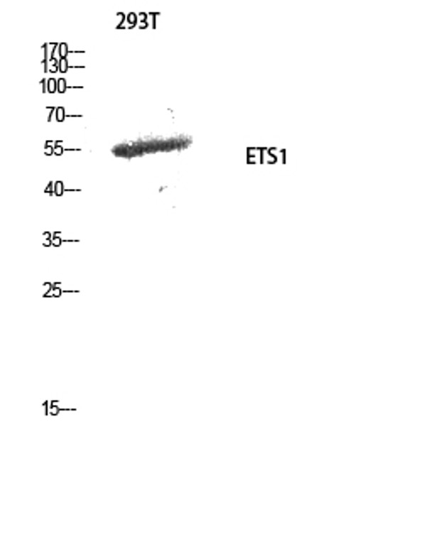
Rabbit polyclonal antibody anti-Protein C-ets-1 (11-60 aa) is suitable for use in Western Blot, Immunohistochemistry, Immunofluorescence and ELISA research applications. |
| Clonality: |
Polyclonal |
| Conjugation: |
Unconjugated |
| Isotype: |
IgG |
| Formulation: |
Liquid in PBS containing 50% Glycerol, 0.5% BSA and 0.02% Sodium Azide. |
| Purification: |
The antibody was affinity-purified from rabbit antiserum by affinity-chromatography using epitope-specific immunogen. |
| Concentration: |
1 mg/mL |
| Dilution Range: |
WB 1:500-1:2000IHC 1:100-1:300IF 1:200-1:1000ELISA 1:5000 |
| Storage Instruction: |
Store at-20°C for up to 1 year from the date of receipt, and avoid repeat freeze-thaw cycles. |
| Gene Symbol: |
ETS1 |
| Gene ID: |
2113 |
| Uniprot ID: |
ETS1_HUMAN |
| Immunogen Region: |
11-60 aa |
| Specificity: |
ETS1 Polyclonal Antibody detects endogenous levels of ETS1 protein. |
| Immunogen: |
The antiserum was produced against synthesized peptide derived from the human ETS1 at the amino acid range 11-60 |
| Post Translational Modifications | Sumoylated on Lys-15 and Lys-227, preferentially with SUMO2.which inhibits transcriptional activity. Ubiquitinated.which induces proteasomal degradation. Phosphorylation at Ser-251, Ser-282 and Ser-285 by CaMK2/CaMKII in response to calcium signaling decreases affinity for DNA: an increasing number of phosphoserines causes DNA-binding to become progressively weaker. |
| Function | Transcription factor. Directly controls the expression of cytokine and chemokine genes in a wide variety of different cellular contexts. May control the differentiation, survival and proliferation of lymphoid cells. May also regulate angiogenesis through regulation of expression of genes controlling endothelial cell migration and invasion. Isoform Ets-1 p27: Acts as a dominant-negative for isoform c-ETS-1A. |
| Protein Name | Protein C-Ets-1P54 |
| Database Links | Reactome: R-HSA-2559585 |
| Cellular Localisation | NucleusCytoplasmDelocalizes From Nucleus To Cytoplasm When Coexpressed With Isoform Ets-1 P27 |
| Alternative Antibody Names | Anti-Protein C-Ets-1 antibodyAnti-P54 antibodyAnti-ETS1 antibodyAnti-EWSR2 antibody |
Information sourced from Uniprot.org
12 months for antibodies. 6 months for ELISA Kits. Please see website T&Cs for further guidance


