| Host: |
Rabbit |
| Applications: |
WB/IHC/IF/ELISA |
| Reactivity: |
Human/Mouse/Rat |
| Note: |
STRICTLY FOR FURTHER SCIENTIFIC RESEARCH USE ONLY (RUO). MUST NOT TO BE USED IN DIAGNOSTIC OR THERAPEUTIC APPLICATIONS. |
| Short Description: |
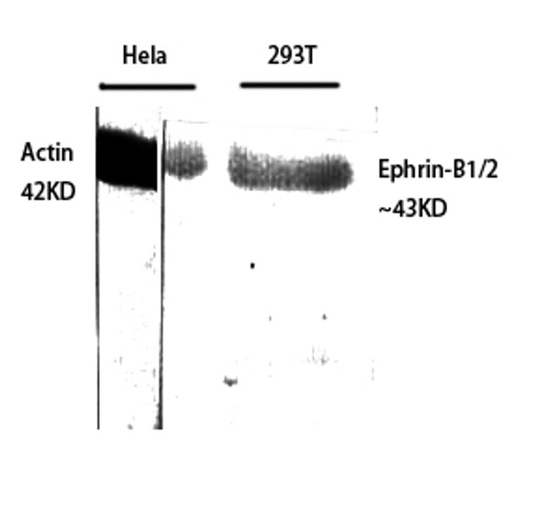
Rabbit polyclonal antibody anti-Ephrin-B1 and Ephrin-B2 (284-333 aa) is suitable for use in Western Blot, Immunohistochemistry, Immunofluorescence and ELISA research applications. |
| Clonality: |
Polyclonal |
| Conjugation: |
Unconjugated |
| Isotype: |
IgG |
| Formulation: |
Liquid in PBS containing 50% Glycerol, 0.5% BSA and 0.02% Sodium Azide. |
| Purification: |
The antibody was affinity-purified from rabbit antiserum by affinity-chromatography using epitope-specific immunogen. |
| Concentration: |
1 mg/mL |
| Dilution Range: |
WB 1:500-1:2000IHC 1:100-1:300ELISA 1:40000IF 1:50-200 |
| Storage Instruction: |
Store at-20°C for up to 1 year from the date of receipt, and avoid repeat freeze-thaw cycles. |
| Gene Symbol: |
EFNB1EFNB2 |
| Gene ID: |
19471948 |
| Uniprot ID: |
EFNB1_HUMANEFNB2_HUMAN |
| Immunogen Region: |
284-333 aa |
| Specificity: |
Ephrin-B1/2 Polyclonal Antibody detects endogenous levels of Ephrin-B1/2 protein. |
| Immunogen: |
The antiserum was produced against synthesized peptide derived from the human EFNB1/2 at the amino acid range 284-333 |
| Post Translational Modifications | Inducible phosphorylation of tyrosine residues in the cytoplasmic domain. Proteolytically processed. The ectodomain is cleaved, probably by a metalloprotease, to produce a membrane-tethered C-terminal fragment. This fragment is then further processed by the gamma-secretase complex to yield a soluble intracellular domain peptide which can translocate to the nucleus. The intracellular domain peptide is highly labile suggesting that it is targeted for degradation by the proteasome. |
| Function | Cell surface transmembrane ligand for Eph receptors, a family of receptor tyrosine kinases which are crucial for migration, repulsion and adhesion during neuronal, vascular and epithelial development. Binding to Eph receptors residing on adjacent cells leads to contact-dependent bidirectional signaling into neighboring cells. Shows high affinity for the receptor tyrosine kinase EPHB1/ELK. Can also bind EPHB2 and EPHB3. Binds to, and induces collapse of, commissural axons/growth cones in vitro. May play a role in constraining the orientation of longitudinally projecting axons. |
| Protein Name | Ephrin-B1Efl-3Elk LigandElk-LEph-Related Receptor Tyrosine Kinase Ligand 2Lerk-2 Cleaved Into - Ephrin-B1 C-Terminal FragmentEphrin-B1 Ctf - Ephrin-B1 Intracellular DomainEphrin-B1 Icd |
| Database Links | Reactome: R-HSA-2682334Reactome: R-HSA-3928662Reactome: R-HSA-3928664Reactome: R-HSA-3928665 |
| Cellular Localisation | Cell MembraneSingle-Pass Type I Membrane ProteinMembrane RaftMay Recruit Grip1 And Grip2 To Membrane Raft DomainsEphrin-B1 C-Terminal Fragment: Cell MembraneEphrin-B1 Intracellular Domain: NucleusColocalizes With Zhx2 In The Nucleus |
| Alternative Antibody Names | Anti-Ephrin-B1 antibodyAnti-Efl-3 antibodyAnti-Elk Ligand antibodyAnti-Elk-L antibodyAnti-Eph-Related Receptor Tyrosine Kinase Ligand 2 antibodyAnti-Lerk-2 Cleaved Into - Ephrin-B1 C-Terminal Fragment antibodyAnti-Ephrin-B1 Ctf - Ephrin-B1 Intracellular Domain antibodyAnti-Ephrin-B1 Icd antibodyAnti-EFNB1 antibodyAnti-EFL3 antibodyAnti-EPLG2 antibodyAnti-LERK2 antibody |
Information sourced from Uniprot.org
12 months for antibodies. 6 months for ELISA Kits. Please see website T&Cs for further guidance


