| Host: |
Rabbit |
| Applications: |
WB/IHC/IF/ELISA |
| Reactivity: |
Human/Mouse/Rat |
| Note: |
STRICTLY FOR FURTHER SCIENTIFIC RESEARCH USE ONLY (RUO). MUST NOT TO BE USED IN DIAGNOSTIC OR THERAPEUTIC APPLICATIONS. |
| Short Description: |
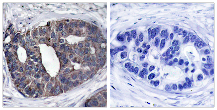
Rabbit polyclonal antibody anti-Docking protein 1 (329-378 aa) is suitable for use in Western Blot, Immunohistochemistry, Immunofluorescence and ELISA research applications. |
| Clonality: |
Polyclonal |
| Conjugation: |
Unconjugated |
| Isotype: |
IgG |
| Formulation: |
Liquid in PBS containing 50% Glycerol, 0.5% BSA and 0.02% Sodium Azide. |
| Purification: |
The antibody was affinity-purified from rabbit antiserum by affinity-chromatography using epitope-specific immunogen. |
| Concentration: |
1 mg/mL |
| Dilution Range: |
WB 1:500-1:2000IHC 1:100-1:300IF 1:200-1:1000ELISA 1:5000 |
| Storage Instruction: |
Store at-20°C for up to 1 year from the date of receipt, and avoid repeat freeze-thaw cycles. |
| Gene Symbol: |
DOK1 |
| Gene ID: |
1796 |
| Uniprot ID: |
DOK1_HUMAN |
| Immunogen Region: |
329-378 aa |
| Specificity: |
Dok-1 Polyclonal Antibody detects endogenous levels of Dok-1 protein. |
| Immunogen: |
The antiserum was produced against synthesized peptide derived from the human p62 Dok at the amino acid range 329-378 |
| Post Translational Modifications | Constitutively tyrosine-phosphorylated. Phosphorylated by TEC. Phosphorylated by LYN. Phosphorylated on tyrosine residues by the insulin receptor kinase. Results in the negative regulation of the insulin signaling pathway. Phosphorylated on tyrosine residues by SRMS. |
| Function | DOK proteins are enzymatically inert adaptor or scaffolding proteins. They provide a docking platform for the assembly of multimolecular signaling complexes. DOK1 appears to be a negative regulator of the insulin signaling pathway. Modulates integrin activation by competing with talin for the same binding site on ITGB3. |
| Protein Name | Docking Protein 1Downstream Of Tyrosine Kinase 1P62(DokPp62 |
| Database Links | Reactome: R-HSA-8849469Reactome: R-HSA-8853659 |
| Cellular Localisation | Isoform 1: CytoplasmNucleusIsoform 3: CytoplasmPerinuclear Region |
| Alternative Antibody Names | Anti-Docking Protein 1 antibodyAnti-Downstream Of Tyrosine Kinase 1 antibodyAnti-P62(Dok antibodyAnti-Pp62 antibodyAnti-DOK1 antibody |
Information sourced from Uniprot.org
12 months for antibodies. 6 months for ELISA Kits. Please see website T&Cs for further guidance


