| Host: |
Rabbit |
| Applications: |
WB/IHC |
| Reactivity: |
Human/Mouse/Rat |
| Note: |
STRICTLY FOR FURTHER SCIENTIFIC RESEARCH USE ONLY (RUO). MUST NOT TO BE USED IN DIAGNOSTIC OR THERAPEUTIC APPLICATIONS. |
| Short Description: |
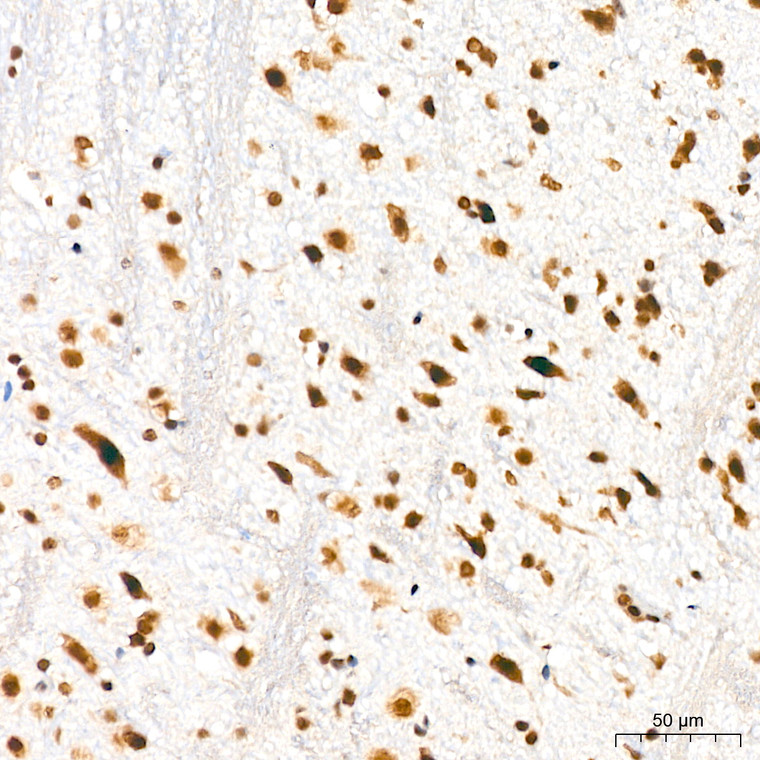
Rabbit monoclonal antibody anti-DKC1 (300-400) is suitable for use in Western Blot and Immunohistochemistry research applications. |
| Clonality: |
Monoclonal |
| Clone ID: |
S3MR |
| Conjugation: |
Unconjugated |
| Isotype: |
IgG |
| Formulation: |
PBS with 0.02% Sodium Azide, 0.05% BSA, 50% Glycerol, pH7.3. |
| Purification: |
Affinity purification |
| Dilution Range: |
WB 1:500-1:2000IHC-P 1:50-1:200 |
| Storage Instruction: |
Store at-20°C for up to 1 year from the date of receipt, and avoid repeat freeze-thaw cycles. |
| Gene Symbol: |
DKC1 |
| Gene ID: |
1736 |
| Uniprot ID: |
DKC1_HUMAN |
| Immunogen Region: |
300-400 |
| Immunogen: |
A synthetic peptide corresponding to a sequence within amino acids 300-400 of human DKC1 (O60832). |
| Immunogen Sequence: |
VMKDSAVNAICYGAKIMLPG VLRYEDGIEVNQEIVVITTK GEAICMAIALMTTAVISTCD HGIVAKIKRVIMERDTYPRK WGLGPKASQKKLMIKQGLLD K |
| Tissue Specificity | Ubiquitously expressed. |
| Function | Isoform 1: Catalytic subunit of H/ACA small nucleolar ribonucleoprotein (H/ACA snoRNP) complex, which catalyzes pseudouridylation of rRNA. This involves the isomerization of uridine such that the ribose is subsequently attached to C5, instead of the normal N1. Each rRNA can contain up to 100 pseudouridine ('psi') residues, which may serve to stabilize the conformation of rRNAs. Required for ribosome biogenesis and telomere maintenance. Also required for correct processing or intranuclear trafficking of TERC, the RNA component of the telomerase reverse transcriptase (TERT) holoenzyme. Isoform 3: Promotes cell to cell and cell to substratum adhesion, increases the cell proliferation rate and leads to cytokeratin hyper-expression. |
| Protein Name | H/Aca Ribonucleoprotein Complex Subunit Dkc1Cbf5 HomologDyskerinNopp140-Associated Protein Of 57 KdaNucleolar Protein Nap57Nucleolar Protein Family A Member 4Snornp Protein Dkc1 |
| Database Links | Reactome: R-HSA-171319Reactome: R-HSA-6790901 |
| Cellular Localisation | Isoform 1: NucleusNucleolusNucleusCajal BodyAlso Localized To Cajal Bodies (Coiled Bodies)Isoform 3: Cytoplasm |
| Alternative Antibody Names | Anti-H/Aca Ribonucleoprotein Complex Subunit Dkc1 antibodyAnti-Cbf5 Homolog antibodyAnti-Dyskerin antibodyAnti-Nopp140-Associated Protein Of 57 Kda antibodyAnti-Nucleolar Protein Nap57 antibodyAnti-Nucleolar Protein Family A Member 4 antibodyAnti-Snornp Protein Dkc1 antibodyAnti-DKC1 antibodyAnti-NOLA4 antibody |
Information sourced from Uniprot.org
12 months for antibodies. 6 months for ELISA Kits. Please see website T&Cs for further guidance


