


Anti-CTNNB1 antibody (C-Term) (STJA0005235)
SPECIFICATIONS
ClonalityPolyclonal
HostRabbit
ConjugationUnconjugated
IsotypeIgG
ImmunogenSynthetic peptide corresponding to amino acid residues from the C-terminal region of rat beta Catenin, conjugated to keyhole limpet hemocyanin (KLH).
General Information
| Short Description | Rabbit polyclonal anti-beta Catenin (C-Term) for use in WB and ICC in Mouse, Rat and Human samples. Datasheet included with dilution recommendations, and related reagents. |
| Applications | WB/ICC |
| Host | Rabbit |
| Reactivity | Mouse/Rat/Human |
| Note | STRICTLY FOR FURTHER SCIENTIFIC RESEARCH USE ONLY (RUO). MUST NOT TO BE USED IN DIAGNOSTIC OR THERAPEUTIC APPLICATIONS. |
Product Properties
| Clonality | Polyclonal |
| Isotype | IgG |
| Conjugation | Unconjugated |
| Purification | Antigen Affinity Purified from Pooled Serum |
| Dilution Range | WB 1:1000ICC 1:200 |
| Formulation | 10 mM HEPES (pH 7.5) , 150 mM NaCl, 100 µg per ml BSA and 50% glycerol. |
| Storage Instruction | Store at-20°C for up to 1 year from the date of receipt, and avoid repeat freeze-thaw cycles. |
Target Information
| Gene Symbol | Ctnnb1 |
| Gene ID | 84353 |
| Uniprot ID | CTNB1_RAT |
| Immunogen | Synthetic peptide corresponding to amino acid residues from the C-terminal region of rat beta Catenin, conjugated to keyhole limpet hemocyanin (KLH). |
| Immunogen Region | C-Term |
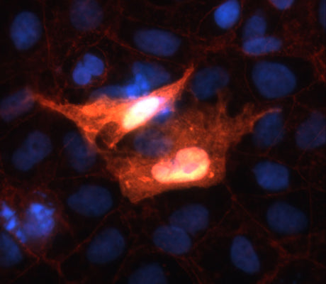
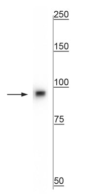
| Specificity | Specific for endogenous levels of the ~ 92 kDa beta Catenin protein. Immunolabeling of the beta Catenin band is completely blocked by preadsorption of the antibody with the peptide used as antigen. |
Additional Info
| Tissue Specificity | Expressed in the testis. |
| Post Translational Modifications | Phosphorylation by GSK3B requires prior phosphorylation of Ser-45 by another kinase. Phosphorylation proceeds then from Thr-41 to Ser-33. Phosphorylated by NEK2. EGF stimulates tyrosine phosphorylation. Phosphorylated on Ser-33 and Ser-37 by HIPK2. This phosphorylation triggers proteasomal degradation. Phosphorylation at Ser-552 by AMPK promotes stabilization of the protein, enhancing TCF/LEF-mediated transcription. Phosphorylation on Ser-191 and Ser-246 by CDK5. Phosphorylation by CDK2 regulates insulin internalization. Phosphorylation by PTK6 at Tyr-64, Tyr-142, Tyr-331 and/or Tyr-333 with the predominant site at Tyr-64 is not essential for inhibition of transcriptional activity. Phosphorylation by SRC at Tyr-333 promotes interaction with isoform M2 of PKM (PKM2).promoting transcription activation. Ubiquitinated by the SCF(BTRC) E3 ligase complex when phosphorylated by GSK3B, leading to its degradation. Ubiquitinated by a E3 ubiquitin ligase complex containing UBE2D1, SIAH1, CACYBP/SIP, SKP1, APC and TBL1X, leading to its subsequent proteasomal degradation. Ubiquitinated and degraded following interaction with SOX9. Ubiquitinated via 'Lys-11'- and 'Lys-29'-linked ubiquitin chains by UBR5, leading to its stabilization. S-nitrosylation at Cys-619 within adherens junctions promotes VEGF-induced, NO-dependent endothelial cell permeability by disrupting interaction with E-cadherin, thus mediating disassembly adherens junctions. O-glycosylation at Ser-23 decreases nuclear localization and transcriptional activity, and increases localization to the plasma membrane and interaction with E-cadherin CDH1. Deacetylated at Lys-49 by SIRT1. |
| Function | Key downstream component of the canonical Wnt signaling pathway. In the absence of Wnt, forms a complex with AXIN1, AXIN2, APC, CSNK1A1 and GSK3B that promotes phosphorylation on N-terminal Ser and Thr residues and ubiquitination of CTNNB1 via BTRC and its subsequent degradation by the proteasome. In the presence of Wnt ligand, CTNNB1 is not ubiquitinated and accumulates in the nucleus, where it acts as a coactivator for transcription factors of the TCF/LEF family, leading to activate Wnt responsive genes. Also acts as a coactivator for other transcription factors, such as NR5A2. Promotes epithelial to mesenchymal transition/mesenchymal to epithelial transition (EMT/MET) via driving transcription of CTNNB1/TCF-target genes. Involved in the regulation of cell adhesion, as component of an E-cadherin:catenin adhesion complex. Acts as a negative regulator of centrosome cohesion. Involved in the CDK2/PTPN6/CTNNB1/CEACAM1 pathway of insulin internalization. Blocks anoikis of malignant kidney and intestinal epithelial cells and promotes their anchorage-independent growth by down-regulating DAPK2. Disrupts PML function and PML-NB formation by inhibiting RANBP2-mediated sumoylation of PML. Promotes neurogenesis by maintaining sympathetic neuroblasts within the cell cycle. Involved in chondrocyte differentiation via interaction with SOX9: SOX9-binding competes with the binding sites of TCF/LEF within CTNNB1, thereby inhibiting the Wnt signaling. Acts as a positive regulator of odontoblast differentiation during mesenchymal tooth germ formation, via promoting the transcription of differentiation factors such as LEF1, BMP2 and BMP4. Activity is repressed in a MSX1-mediated manner at the bell stage of mesenchymal tooth germ formation which prevents premature differentiation of odontoblasts. |
| Protein Name | Catenin Beta-1Beta-Catenin |
| Database Links | Reactome: R-RNO-195253Reactome: -RNO-196299Reactome: -RNO-201681Reactome: -RNO-201722Reactome: -RNO-3134973Reactome: -RNO-351906Reactome: -RNO-3769402Reactome: -RNO-4086398Reactome: -RNO-4641262Reactome: -RNO-5218920Reactome: -RNO-525793Reactome: -RNO-8951430Reactome: -RNO-9762292Reactome: -RNO-9825892 |
| Cellular Localisation | CytoplasmNucleusCytoskeletonCell JunctionAdherens JunctionCell MembraneMicrotubule Organizing CenterCentrosomeSpindle PoleSynapseCilium Basal BodyColocalized With Rapgef2 And Tjp1 At Cell-Cell ContactsCytoplasmic When It Is Un-Stable (Highly Phosphorylated) Or Bound To Cdh1Translocates To The Nucleus When It Is Stabilized (Low Level Of Phosphorylation)Interaction With Glis2 And Muc1 Promotes Nuclear TranslocationInteraction With Emd Inhibits Nuclear LocalizationThe Majority Of Ctnnb1 Is Localized To The Cell MembraneIn InterphaseColocalizes With Crocc Between Cep250 Puncta At The Proximal End Of CentriolesAnd This Localization Is Dependent On Crocc And Cep250In MitosisWhen Nek2 Activity IncreasesIt Localizes To Centrosomes At Spindle Poles Independent Of CroccColocalizes With Cdk5 In The Cell-Cell Contacts And Plasma Membrane Of Undifferentiated And Differentiated Neuroblastoma CellsInteraction With Fam53b Promotes Translocation To The NucleusTranslocates To The Nucleus In The Presence Of Snail1Ca(2+)-Mediated Localization To The Cell Membrane In Dental Epithelial Cells Is Inhibited Via Wnt3aLocalizes To Cell-Cell Contacts As Keratinocyte Differentiation Progresses |
| Alternative Antibody Names | Anti-Catenin Beta-1 antibodyAnti-Beta-Catenin antibodyAnti-Ctnnb1 antibodyAnti-Catnb antibody |
Information sourced from Uniprot.org