| Host: |
Goat |
| Applications: |
Pep-ELISA/WB/IHC/FC |
| Reactivity: |
Human |
| Note: |
STRICTLY FOR FURTHER SCIENTIFIC RESEARCH USE ONLY (RUO). MUST NOT TO BE USED IN DIAGNOSTIC OR THERAPEUTIC APPLICATIONS. |
| Short Description: |
Goat polyclonal antibody anti-Complement factor H (Internal) is suitable for use in ELISA, Western Blot, Immunohistochemistry and Flow Cytometry research applications. |
| Clonality: |
Polyclonal |
| Conjugation: |
Unconjugated |
| Isotype: |
IgG |
| Formulation: |
0.5 mg/ml in Tris saline, 0.02% sodium azide, pH7.3 with 0.5% bovine serum albumin. NA |
| Purification: |
Purified from goat serum by ammonium sulphate precipitation followed by antigen affinity chromatography using the immunizing peptide. |
| Concentration: |
0.5 mg/mL |
| Dilution Range: |
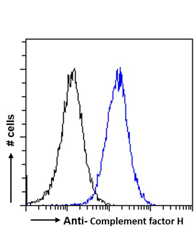
WB-0.03-0.3µg/mlIHC-5µg/mlFC-Flow cytometric analysis of HepG2 cells. 10ug/mlELISA-antibody detection limit dilution 1:32000. |
| Storage Instruction: |
Store at-20°C on receipt and minimise freeze-thaw cycles. |
| Gene Symbol: |
CFH |
| Gene ID: |
3075 |
| Uniprot ID: |
CFAH_HUMAN |
| Immunogen Region: |
Internal |
| Accession Number: |
NP_000177.2 |
| Specificity: |
This antibody is expected to recognize isoform a (NP_000177.2) only. |
| Immunogen Sequence: |
HLVPDRKKDQYK |
| Post Translational Modifications | Sulfated on tyrosine residues. |
| Function | Glycoprotein that plays an essential role in maintaining a well-balanced immune response by modulating complement activation. Acts as a soluble inhibitor of complement, where its binding to self markers such as glycan structures prevents complement activation and amplification on cell surfaces. Accelerates the decay of the complement alternative pathway (AP) C3 convertase C3bBb, thus preventing local formation of more C3b, the central player of the complement amplification loop. As a cofactor of the serine protease factor I, CFH also regulates proteolytic degradation of already-deposited C3b. In addition, mediates several cellular responses through interaction with specific receptors. For example, interacts with CR3/ITGAM receptor and thereby mediates the adhesion of human neutrophils to different pathogens. In turn, these pathogens are phagocytosed and destroyed. (Microbial infection) In the mosquito midgut, binds to the surface of parasite P.falciparum gametocytes and protects the parasite from alternative complement pathway-mediated elimination. |
| Protein Name | Complement Factor HH Factor 1 |
| Database Links | Reactome: R-HSA-977606 |
| Cellular Localisation | Secreted(Microbial Infection) In The Mosquito MidgutLocalizes To PFalciparum (Nf54 Strain) Macrogamete And Young Zygote Cell Membranes |
| Alternative Antibody Names | Anti-Complement Factor H antibodyAnti-H Factor 1 antibodyAnti-CFH antibodyAnti-HF antibodyAnti-HF1 antibodyAnti-HF2 antibody |
Information sourced from Uniprot.org
12 months for antibodies. 6 months for ELISA Kits. Please see website T&Cs for further guidance


