| Host: |
Rabbit |
| Applications: |
WB/IHC/IF |
| Reactivity: |
Human/Mouse/Rat |
| Note: |
STRICTLY FOR FURTHER SCIENTIFIC RESEARCH USE ONLY (RUO). MUST NOT TO BE USED IN DIAGNOSTIC OR THERAPEUTIC APPLICATIONS. |
| Short Description: |
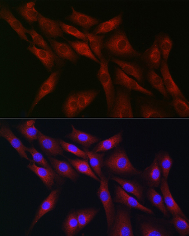
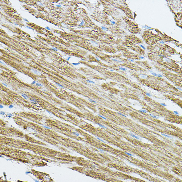
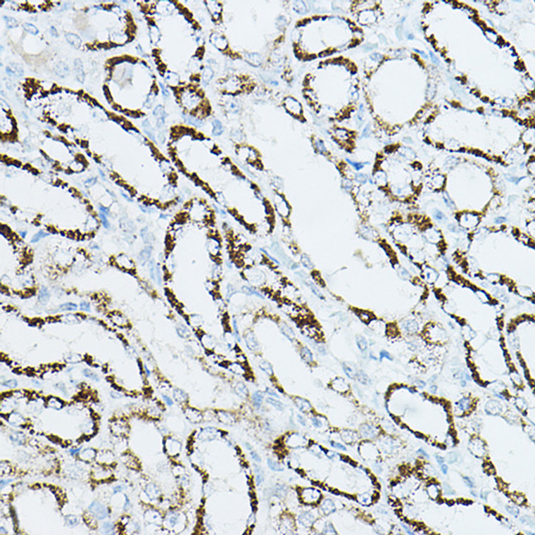
Rabbit polyclonal antibody anti-CDC42 (118-191) is suitable for use in Western Blot, Immunohistochemistry and Immunofluorescence research applications. |
| Clonality: |
Polyclonal |
| Conjugation: |
Unconjugated |
| Isotype: |
IgG |
| Formulation: |
PBS with 0.01% Thimerosal, 50% Glycerol, pH7.3. |
| Purification: |
Affinity purification |
| Dilution Range: |
WB 1:500-1:1000IHC-P 1:50-1:200IF/ICC 1:50-1:200 |
| Storage Instruction: |
Store at-20°C for up to 1 year from the date of receipt, and avoid repeat freeze-thaw cycles. |
| Gene Symbol: |
CDC42 |
| Gene ID: |
998 |
| Uniprot ID: |
CDC42_HUMAN |
| Immunogen Region: |
118-191 |
| Immunogen: |
Recombinant fusion protein containing a sequence corresponding to amino acids 118-191 of human CDC42 (NP_001782.1). |
| Immunogen Sequence: |
DLRDDPSTIEKLAKNKQKPI TPETAEKLARDLKAVKYVEC SALTQKGLKNVFDEAILAAL EPPEPKKSRRCVLL |
| Post Translational Modifications | (Microbial infection) AMPylation at Tyr-32 and Thr-35 are mediated by bacterial enzymes in case of infection by H.somnus and V.parahaemolyticus, respectively. AMPylation occurs in the effector region and leads to inactivation of the GTPase activity by preventing the interaction with downstream effectors, thereby inhibiting actin assembly in infected cells. It is unclear whether some human enzyme mediates AMPylation.FICD has such ability in vitro but additional experiments remain to be done to confirm results in vivo. Phosphorylated by SRC in an EGF-dependent manner, this stimulates the binding of the Rho-GDP dissociation inhibitor RhoGDI. (Microbial infection) Glycosylated at Tyr-32 by Photorhabdus asymbiotica toxin PAU_02230. Mono-O-GlcNAcylation by PAU_02230 inhibits downstream signaling by an impaired interaction with diverse regulator and effector proteins of CDC42 and leads to actin disassembly. (Microbial infection) Glucosylated at Thr-35 by C.difficile toxins TcdA and TcdB in the colonic epithelium. Monoglucosylation completely prevents the recognition of the downstream effector, blocking the GTPases in their inactive form, leading to actin cytoskeleton disruption and cell death, resulting in the loss of colonic epithelial barrier function. (Microbial infection) Glycosylated (O-GlcNAcylated) at Thr-35 by C.novyi toxin TcdA. O-GlcNAcylation completely prevents the recognition of the downstream effector, blocking the GTPases in their inactive form, leading to actin cytoskeleton disruption. |
| Function | Plasma membrane-associated small GTPase which cycles between an active GTP-bound and an inactive GDP-bound state. In active state binds to a variety of effector proteins to regulate cellular responses. Involved in epithelial cell polarization processes. Regulates the bipolar attachment of spindle microtubules to kinetochores before chromosome congression in metaphase. Regulates cell migration. In neurons, plays a role in the extension and maintenance of the formation of filopodia, thin and actin-rich surface projections. Required for DOCK10-mediated spine formation in Purkinje cells and hippocampal neurons. In podocytes, facilitates filopodia and podosomes formation upon DOCK11-activation. Upon activation by CaMKII, modulates dendritic spine structural plasticity by relaying CaMKII transient activation to synapse-specific, long-term signaling. Also plays a role in phagocytosis through organization of the F-actin cytoskeleton associated with forming phagocytic cups. |
| Protein Name | Cell Division Control Protein 42 HomologG25k Gtp-Binding Protein |
| Database Links | Reactome: R-HSA-114604Reactome: R-HSA-182971Reactome: R-HSA-2029482Reactome: R-HSA-389359Reactome: R-HSA-3928662Reactome: R-HSA-418885Reactome: R-HSA-428543Reactome: R-HSA-4420097Reactome: R-HSA-525793Reactome: R-HSA-5625970Reactome: R-HSA-5626467Reactome: R-HSA-5627123Reactome: R-HSA-5663213Reactome: R-HSA-5663220Reactome: R-HSA-5687128Reactome: R-HSA-8950505Reactome: R-HSA-8964616Reactome: R-HSA-9013148Reactome: R-HSA-9013149Reactome: R-HSA-9013404Reactome: R-HSA-9013406Reactome: R-HSA-9013408Reactome: R-HSA-9013409Reactome: R-HSA-9013420Reactome: R-HSA-9013423Reactome: R-HSA-9013424Reactome: R-HSA-9664422Reactome: R-HSA-983231 |
| Cellular Localisation | Cell MembraneLipid-AnchorCytoplasmic SideCytoplasmCytoskeletonMicrotubule Organizing CenterCentrosomeSpindleMidbodyCell ProjectionDendriteLocalizes To Spindle During Prometaphase CellsMoves To The Central Spindle As Cells Progressed Through Anaphase To TelophaseLocalizes At The End Of Cytokinesis In The Intercellular Bridge Formed Between Two Daughter CellsIts Localization Is Regulated By The Activities Of Guanine Nucleotide Exchange Factor Ect2 And Gtpase Activating Protein Racgap1Colocalizes With Nek6 In The CentrosomeIn Its Active Gtp-Bound Form Localizes To The Leading Edge Membrane Of Migrating Dendritic Cells |
| Alternative Antibody Names | Anti-Cell Division Control Protein 42 Homolog antibodyAnti-G25k Gtp-Binding Protein antibodyAnti-CDC42 antibody |
Information sourced from Uniprot.org
12 months for antibodies. 6 months for ELISA Kits. Please see website T&Cs for further guidance


