| Host: |
Rabbit |
| Applications: |
WB/IF/ELISA |
| Reactivity: |
Human/Rat/Mouse |
| Note: |
STRICTLY FOR FURTHER SCIENTIFIC RESEARCH USE ONLY (RUO). MUST NOT TO BE USED IN DIAGNOSTIC OR THERAPEUTIC APPLICATIONS. |
| Short Description: |
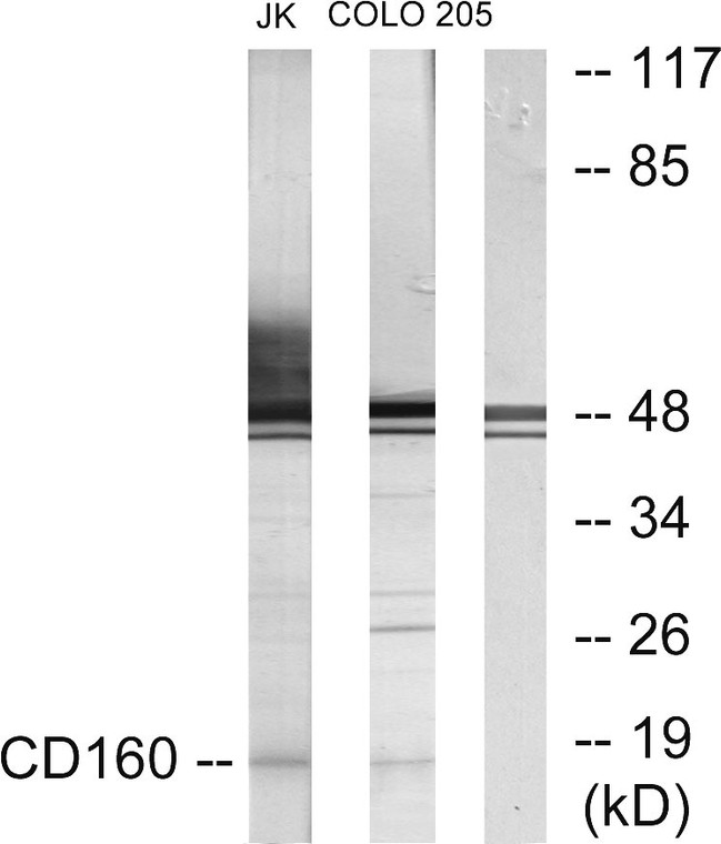
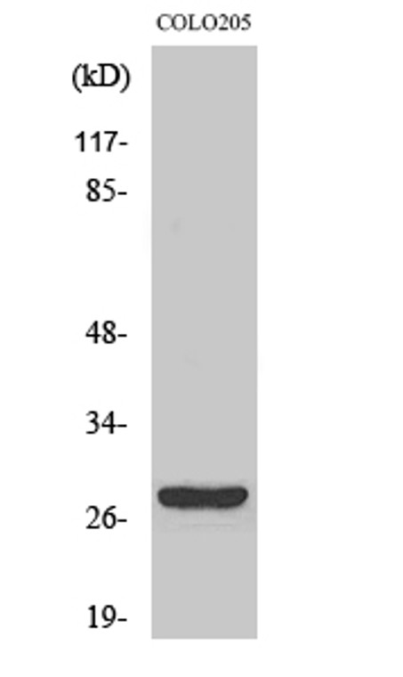
Rabbit polyclonal antibody anti-CD160 antigen (21-70 aa) is suitable for use in Western Blot, Immunofluorescence and ELISA research applications. |
| Clonality: |
Polyclonal |
| Conjugation: |
Unconjugated |
| Isotype: |
IgG |
| Formulation: |
Liquid in PBS containing 50% Glycerol, 0.5% BSA and 0.02% Sodium Azide. |
| Purification: |
The antibody was affinity-purified from rabbit antiserum by affinity-chromatography using epitope-specific immunogen. |
| Concentration: |
1 mg/mL |
| Dilution Range: |
WB 1:500-1:2000IF 1:200-1:1000ELISA 1:20000 |
| Storage Instruction: |
Store at-20°C for up to 1 year from the date of receipt, and avoid repeat freeze-thaw cycles. |
| Gene Symbol: |
CD160 |
| Gene ID: |
11126 |
| Uniprot ID: |
BY55_HUMAN |
| Immunogen Region: |
21-70 aa |
| Specificity: |
CD160 Polyclonal Antibody detects endogenous levels of CD160 protein. |
| Immunogen: |
The antiserum was produced against synthesized peptide derived from the human CD160 at the amino acid range 21-70 |
| Function | CD160 antigen: Receptor on immune cells capable to deliver stimulatory or inhibitory signals that regulate cell activation and differentiation. Exists as a GPI-anchored and as a transmembrane form, each likely initiating distinct signaling pathways via phosphoinositol 3-kinase in activated NK cells and via LCK and CD247/CD3 zeta chain in activated T cells. Receptor for both classical and non-classical MHC class I molecules. In the context of acute viral infection, recognizes HLA-C and triggers NK cell cytotoxic activity, likely playing a role in anti-viral innate immune response. On CD8+ T cells, binds HLA-A2-B2M in complex with a viral peptide and provides a costimulatory signal to activated/memory T cells. Upon persistent antigen stimulation, such as occurs during chronic viral infection, may progressively inhibit TCR signaling in memory CD8+ T cells, contributing to T cell exhaustion. On endothelial cells, recognizes HLA-G and controls angiogenesis in immune privileged sites. Receptor or ligand for TNF superfamily member TNFRSF14, participating in bidirectional cell-cell contact signaling between antigen presenting cells and lymphocytes. Upon ligation of TNFRSF14, provides stimulatory signal to NK cells enhancing IFNG production and anti-tumor immune response. On activated CD4+ T cells, interacts with TNFRSF14 and down-regulates CD28 costimulatory signaling, restricting memory and alloantigen-specific immune response. In the context of bacterial infection, acts as a ligand for TNFRSF14 on epithelial cells, triggering the production of antimicrobial proteins and pro-inflammatory cytokines. CD160 antigen, soluble form: The soluble GPI-cleaved form, usually released by activated lymphocytes, might play an immune regulatory role by limiting lymphocyte effector functions. |
| Protein Name | Cd160 AntigenNatural Killer Cell Receptor By55Cd Antigen Cd160 Cleaved Into - Cd160 Antigen - Soluble Form |
| Database Links | Reactome: R-HSA-198933 |
| Cellular Localisation | Cd160 Antigen: Cell MembraneLipid-AnchorGpi-AnchorCd160 AntigenSoluble Form: SecretedReleased From The Cell Membrane By Gpi Cleavage |
| Alternative Antibody Names | Anti-Cd160 Antigen antibodyAnti-Natural Killer Cell Receptor By55 antibodyAnti-Cd Antigen Cd160 Cleaved Into - Cd160 Antigen - Soluble Form antibodyAnti-CD160 antibodyAnti-BY55 antibody |
Information sourced from Uniprot.org
12 months for antibodies. 6 months for ELISA Kits. Please see website T&Cs for further guidance


