| Host: |
Rabbit |
| Applications: |
WB/IHC/IF |
| Reactivity: |
Human/Mouse/Rat |
| Note: |
STRICTLY FOR FURTHER SCIENTIFIC RESEARCH USE ONLY (RUO). MUST NOT TO BE USED IN DIAGNOSTIC OR THERAPEUTIC APPLICATIONS. |
| Short Description: |
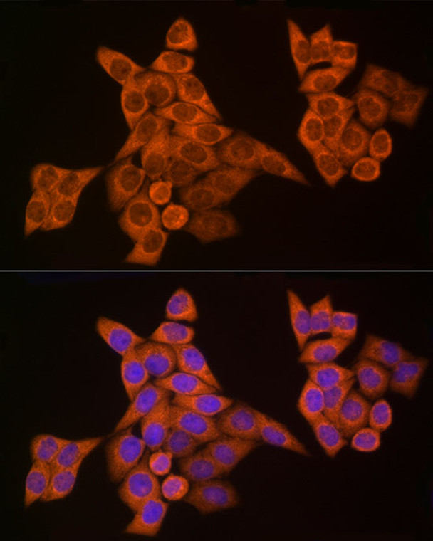
Rabbit polyclonal antibody anti-Caspase-8 (376-479) is suitable for use in Western Blot, Immunohistochemistry and Immunofluorescence research applications. |
| Clonality: |
Polyclonal |
| Conjugation: |
Unconjugated |
| Isotype: |
IgG |
| Formulation: |
PBS with 0.05% Proclin300, 50% Glycerol, pH7.3. |
| Purification: |
Affinity purification |
| Dilution Range: |
WB 1:500-1:1000IHC-P 1:50-1:200IF/ICC 1:50-1:200 |
| Storage Instruction: |
Store at-20°C for up to 1 year from the date of receipt, and avoid repeat freeze-thaw cycles. |
| Gene Symbol: |
CASP8 |
| Gene ID: |
841 |
| Uniprot ID: |
CASP8_HUMAN |
| Immunogen Region: |
376-479 |
| Immunogen: |
A synthetic peptide corresponding to a sequence within amino acids 376-479 of human Caspase 8 (NP_203519.1). |
| Immunogen Sequence: |
EEQPYLEMDLSSPQTRYIPD EADFLLGMATVNNCVSYRNP AEGTWYIQSLCQSLRERCPR GDDILTILTEVNYEVSNKDD KKNMGKQMPQPTFTLRKKLV FPSD |
| Tissue Specificity | Isoform 1, isoform 5 and isoform 7 are expressed in a wide variety of tissues. Highest expression in peripheral blood leukocytes, spleen, thymus and liver. Barely detectable in brain, testis and skeletal muscle. |
| Post Translational Modifications | (Microbial infection) Proteolytically cleaved by the cowpox virus CRMA death inhibitory protein. Generation of the subunits requires association with the death-inducing signaling complex (DISC), whereas additional processing is likely due to the autocatalytic activity of the activated protease. GZMB and CASP10 can be involved in these processing events. Phosphorylation on Ser-387 during mitosis by CDK1 inhibits activation by proteolysis and prevents apoptosis. This phosphorylation occurs in cancer cell lines, as well as in primary breast tissues and lymphocytes. (Microbial infection) ADP-riboxanation by C.violaceum CopC blocks CASP8 processing, preventing CASP8 activation and ability to mediate extrinsic apoptosis. |
| Function | Thiol protease that plays a key role in programmed cell death by acting as a molecular switch for apoptosis, necroptosis and pyroptosis, and is required to prevent tissue damage during embryonic development and adulthood. Initiator protease that induces extrinsic apoptosis by mediating cleavage and activation of effector caspases responsible for the TNFRSF6/FAS mediated and TNFRSF1A induced cell death. Cleaves and activates effector caspases CASP3, CASP4, CASP6, CASP7, CASP9 and CASP10. Binding to the adapter molecule FADD recruits it to either receptor TNFRSF6/FAS mediated or TNFRSF1A. The resulting aggregate called death-inducing signaling complex (DISC) performs CASP8 proteolytic activation. The active dimeric enzyme is then liberated from the DISC and free to activate downstream apoptotic proteases. Proteolytic fragments of the N-terminal propeptide (termed CAP3, CAP5 and CAP6) are likely retained in the DISC. In addition to extrinsic apoptosis, also acts as a negative regulator of necroptosis: acts by cleaving RIPK1 at 'Asp-324', which is crucial to inhibit RIPK1 kinase activity, limiting TNF-induced apoptosis, necroptosis and inflammatory response. Also able to initiate pyroptosis by mediating cleavage and activation of gasdermin-C and -D (GSDMC and GSDMD, respectively): gasdermin cleavage promotes release of the N-terminal moiety that binds to membranes and forms pores, triggering pyroptosis. Initiates pyroptosis following inactivation of MAP3K7/TAK1. Also acts as a regulator of innate immunity by mediating cleavage and inactivation of N4BP1 downstream of TLR3 or TLR4, thereby promoting cytokine production. May participate in the Granzyme B (GZMB) cell death pathways. Cleaves PARP1 and PARP2. Isoform 5: Lacks the catalytic site and may interfere with the pro-apoptotic activity of the complex. Isoform 6: Lacks the catalytic site and may interfere with the pro-apoptotic activity of the complex. Isoform 7: Lacks the catalytic site and may interfere with the pro-apoptotic activity of the complex (Probable). Acts as an inhibitor of the caspase cascade. Isoform 8: Lacks the catalytic site and may interfere with the pro-apoptotic activity of the complex. |
| Protein Name | Caspase-8Casp-8Apoptotic Cysteine ProteaseApoptotic Protease Mch-5Cap4Fadd-Homologous Ice/Ced-3-Like ProteaseFadd-Like IceFliceIce-Like Apoptotic Protease 5Mort1-Associated Ced-3 HomologMach Cleaved Into - Caspase-8 Subunit P18 - Caspase-8 Subunit P10 |
| Database Links | Reactome: R-HSA-111465Reactome: R-HSA-140534Reactome: R-HSA-168638Reactome: R-HSA-2562578Reactome: R-HSA-264870Reactome: R-HSA-3371378Reactome: R-HSA-5213460Reactome: R-HSA-5218900Reactome: R-HSA-5357786Reactome: R-HSA-5357905Reactome: R-HSA-5660668Reactome: R-HSA-5675482Reactome: R-HSA-69416Reactome: R-HSA-75108Reactome: R-HSA-75153Reactome: R-HSA-75157Reactome: R-HSA-75158Reactome: R-HSA-9013957Reactome: R-HSA-933543Reactome: R-HSA-9686347Reactome: R-HSA-9693928Reactome: R-HSA-9758274 |
| Cellular Localisation | CytoplasmNucleus |
| Alternative Antibody Names | Anti-Caspase-8 antibodyAnti-Casp-8 antibodyAnti-Apoptotic Cysteine Protease antibodyAnti-Apoptotic Protease Mch-5 antibodyAnti-Cap4 antibodyAnti-Fadd-Homologous Ice/Ced-3-Like Protease antibodyAnti-Fadd-Like Ice antibodyAnti-Flice antibodyAnti-Ice-Like Apoptotic Protease 5 antibodyAnti-Mort1-Associated Ced-3 Homolog antibodyAnti-Mach Cleaved Into - Caspase-8 Subunit P18 - Caspase-8 Subunit P10 antibodyAnti-CASP8 antibodyAnti-MCH5 antibody |
Information sourced from Uniprot.org
12 months for antibodies. 6 months for ELISA Kits. Please see website T&Cs for further guidance


