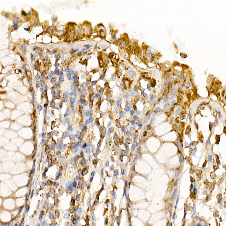
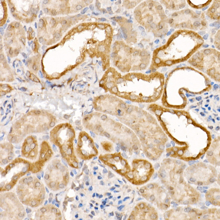
| Tissue Specificity | Highly expressed in lung, skeletal muscle, liver, kidney, spleen and heart, and moderately in testis. No expression in the brain. |
| Post Translational Modifications | Cleavage by different proteases, such as granzyme B (GZMB), caspase-1 (CASP1), caspase-8 (CASP8), caspase-9 (CASP9) or caspase-10 (CASP10) generate the two active subunits. Its involvement in different programmed cell death processes is probably specified by the protease that activates CASP7. Cleaved and activated by initiator caspases (CASP8, CASP9 and/or CASP10), leading to execution phase of apoptosis. Cleavage and maturation by GZMB regulates granzyme-mediated programmed cell death. Cleaved and activated by CASP1 in response to bacterial infection. Propeptide domains can also be cleaved efficiently by CASP3. Active heterodimers between the small subunit of caspase-7 and the large subunit of CASP3, and vice versa, also occur. Also cleaved at the N-terminus at alternative sites by CAPN1, leading to its activation. Phosphorylation at Ser-30 and Ser-239 by PAK2 inhibits its activity. Phosphorylation at Ser-30 prevents cleavage and activation by initiator caspase CASP9, while phosphorylation at Ser-239 prevents thiol protease activity by preventing substrate-binding. (Microbial infection) ADP-riboxanation by C.violaceum CopC blocks CASP7 processing, preventing CASP7 activation and ability to recognize and cleave substrates. |
| Function | Thiol protease involved in different programmed cell death processes, such as apoptosis, pyroptosis or granzyme-mediated programmed cell death, by proteolytically cleaving target proteins. Has a marked preference for Asp-Glu-Val-Asp (DEVD) consensus sequences, with some plasticity for alternate non-canonical sequences. Its involvement in the different programmed cell death processes is probably determined by upstream proteases that activate CASP7. Acts as an effector caspase involved in the execution phase of apoptosis: following cleavage and activation by initiator caspases (CASP8, CASP9 and/or CASP10), mediates execution of apoptosis by catalyzing cleavage of proteins, such as CLSPN, PARP1, PTGES3 and YY1. Compared to CASP3, acts as a minor executioner caspase and cleaves a limited set of target proteins. Acts as a key regulator of the inflammatory response in response to bacterial infection by catalyzing cleavage and activation of the sphingomyelin phosphodiesterase SMPD1 in the extracellular milieu, thereby promoting membrane repair. Regulates pyroptosis in intestinal epithelial cells: cleaved and activated by CASP1 in response to S.typhimurium infection, promoting its secretion to the extracellular milieu, where it catalyzes activation of SMPD1, generating ceramides that repair membranes and counteract the action of gasdermin-D (GSDMD) pores. Regulates granzyme-mediated programmed cell death in hepatocytes: cleaved and activated by granzyme B (GZMB) in response to bacterial infection, promoting its secretion to the extracellular milieu, where it catalyzes activation of SMPD1, generating ceramides that repair membranes and counteract the action of perforin (PRF1) pores. Following cleavage by CASP1 in response to inflammasome activation, catalyzes processing and inactivation of PARP1, alleviating the transcription repressor activity of PARP1. Acts as an inhibitor of type I interferon production during virus-induced apoptosis by mediating cleavage of antiviral proteins CGAS, IRF3 and MAVS, thereby preventing cytokine overproduction. Cleaves and activates sterol regulatory element binding proteins (SREBPs). Cleaves phospholipid scramblase proteins XKR4, XKR8 and XKR9. In case of infection, catalyzes cleavage of Kaposi sarcoma-associated herpesvirus protein ORF57, thereby preventing expression of viral lytic genes. Isoform Beta: Lacks enzymatic activity. |
| Protein Name | Caspase-7Casp-7Apoptotic Protease Mch-3Cmh-1Ice-Like Apoptotic Protease 3Ice-Lap3 Cleaved Into - Caspase-7 Subunit P20 - Caspase-7 Subunit P11 |
| Database Links | Reactome: R-HSA-111459Reactome: R-HSA-111463Reactome: R-HSA-111464Reactome: R-HSA-111465Reactome: R-HSA-111469Reactome: R-HSA-264870 |
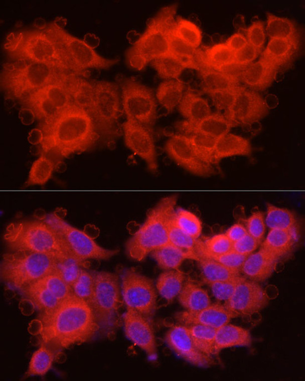
| Cellular Localisation | CytoplasmCytosolNucleusSecretedExtracellular SpaceFollowing Cleavage And Activation By Casp1 Or Granzyme B (Gzmb)Secreted Into The Extracellular Milieu By Passing Through The Gasdermin-D (Gsdmd) Pores Or Perforin (Prf1) PoreRespectively |
| Alternative Antibody Names | Anti-Caspase-7 antibodyAnti-Casp-7 antibodyAnti-Apoptotic Protease Mch-3 antibodyAnti-Cmh-1 antibodyAnti-Ice-Like Apoptotic Protease 3 antibodyAnti-Ice-Lap3 Cleaved Into - Caspase-7 Subunit P20 - Caspase-7 Subunit P11 antibodyAnti-CASP7 antibodyAnti-MCH3 antibody |


