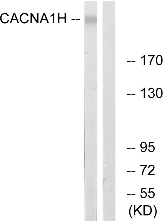
| Host: |
Rabbit |
| Applications: |
WB/ELISA |
| Reactivity: |
Human/Mouse/Rat |
| Note: |
STRICTLY FOR FURTHER SCIENTIFIC RESEARCH USE ONLY (RUO). MUST NOT TO BE USED IN DIAGNOSTIC OR THERAPEUTIC APPLICATIONS. |
| Short Description: |
Rabbit polyclonal antibody anti-Voltage-dependent T-type calcium channel subunit alpha-1H (462-511 aa) is suitable for use in Western Blot and ELISA research applications. |
| Clonality: |
Polyclonal |
| Conjugation: |
Unconjugated |
| Isotype: |
IgG |
| Formulation: |
Liquid in PBS containing 50% Glycerol, 0.5% BSA and 0.02% Sodium Azide. |
| Purification: |
The antibody was affinity-purified from rabbit antiserum by affinity-chromatography using epitope-specific immunogen. |
| Concentration: |
1 mg/mL |
| Dilution Range: |
WB 1:500-1:2000ELISA 1:10000 |
| Storage Instruction: |
Store at-20°C for up to 1 year from the date of receipt, and avoid repeat freeze-thaw cycles. |
| Gene Symbol: |
CACNA1H |
| Gene ID: |
8912 |
| Uniprot ID: |
CAC1H_HUMAN |
| Immunogen Region: |
462-511 aa |
| Specificity: |
T-type Ca++ CP Alpha 1H Polyclonal Antibody detects endogenous levels of T-type Ca++ CP Alpha 1H protein. |
| Immunogen: |
The antiserum was produced against synthesized peptide derived from the human CACNA1H at the amino acid range 462-511 |
| Post Translational Modifications | In response to raising of intracellular calcium, the T-type channels are activated by CaM-kinase II. |
| Function | Voltage-sensitive calcium channel that gives rise to T-type calcium currents. T-type calcium channels belong to the 'low-voltage activated (LVA)' group. A particularity of this type of channel is an opening at quite negative potentials, and a voltage-dependent inactivation. T-type channels serve pacemaking functions in both central neurons and cardiac nodal cells and support calcium signaling in secretory cells and vascular smooth muscle (Probable). They may also be involved in the modulation of firing patterns of neurons. In the adrenal zona glomerulosa, participates in the signaling pathway leading to aldosterone production in response to either AGT/angiotensin II, or hyperkalemia. |
| Protein Name | Voltage-Dependent T-Type Calcium Channel Subunit Alpha-1hLow-Voltage-Activated Calcium Channel Alpha1 3.2 SubunitVoltage-Gated Calcium Channel Subunit Alpha Cav3.2 |
| Database Links | Reactome: R-HSA-419037Reactome: R-HSA-445355 |
| Cellular Localisation | Cell MembraneMulti-Pass Membrane ProteinInteraction With Stac Increases Expression At The Cell Membrane |
| Alternative Antibody Names | Anti-Voltage-Dependent T-Type Calcium Channel Subunit Alpha-1h antibodyAnti-Low-Voltage-Activated Calcium Channel Alpha1 3.2 Subunit antibodyAnti-Voltage-Gated Calcium Channel Subunit Alpha Cav3.2 antibodyAnti-CACNA1H antibody |
Information sourced from Uniprot.org
12 months for antibodies. 6 months for ELISA Kits. Please see website T&Cs for further guidance


