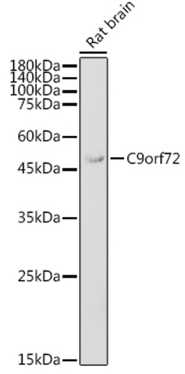
| Host: |
Rabbit |
| Applications: |
WB/IHC/IF |
| Reactivity: |
Human/Mouse/Rat |
| Note: |
STRICTLY FOR FURTHER SCIENTIFIC RESEARCH USE ONLY (RUO). MUST NOT TO BE USED IN DIAGNOSTIC OR THERAPEUTIC APPLICATIONS. |
| Short Description: |
Rabbit polyclonal antibody anti-C9orf72 (9-200) is suitable for use in Western Blot, Immunohistochemistry and Immunofluorescence research applications. |
| Clonality: |
Polyclonal |
| Conjugation: |
Unconjugated |
| Isotype: |
IgG |
| Formulation: |
PBS with 0.01% Thimerosal, 50% Glycerol, pH7.3. |
| Purification: |
Affinity purification |
| Dilution Range: |
WB 1:500-1:1000IHC-P 1:50-1:200IF/ICC 1:50-1:200 |
| Storage Instruction: |
Store at-20°C for up to 1 year from the date of receipt, and avoid repeat freeze-thaw cycles. |
| Gene Symbol: |
C9orf72 |
| Gene ID: |
203228 |
| Uniprot ID: |
CI072_HUMAN |
| Immunogen Region: |
9-200 |
| Immunogen: |
Recombinant fusion protein containing a sequence corresponding to amino acids 9-200 of human C9orf72 (NP_001242983.1). |
| Immunogen Sequence: |
SPAVAKTEIALSGKSPLLAA TFAYWDNILGPRVRHIWAPK TEQVLLSDGEITFLANHTLN GEILRNAESGAIDVKFFVLS EKGVIIVSLIFDGNWNGDRS TYGLSIILPQTELSFYLPLH RVCVDRLTHIIRKGRIWMHK ERQENVQKIILEGTERMEDQ GQSIIPMLTGEVIPVMELLS SMKSHSVPEEID |
| Tissue Specificity | Both isoforms are widely expressed, including kidney, lung, liver, heart, testis and several brain regions, such as cerebellum. Also expressed in the frontal cortex and in lymphoblasts (at protein level). |
| Function | Component of the C9orf72-SMCR8 complex, a complex that has guanine nucleotide exchange factor (GEF) activity and regulates autophagy. In the complex, C9orf72 and SMCR8 probably constitute the catalytic subunits that promote the exchange of GDP to GTP, converting inactive GDP-bound RAB8A and RAB39B into their active GTP-bound form, thereby promoting autophagosome maturation. The C9orf72-SMCR8 complex also acts as a regulator of autophagy initiation by interacting with the ULK1/ATG1 kinase complex and modulating its protein kinase activity. As part of the C9orf72-SMCR8 complex, stimulates RAB8A and RAB11A GTPase activity in vitro. Positively regulates initiation of autophagy by regulating the RAB1A-dependent trafficking of the ULK1/ATG1 kinase complex to the phagophore which leads to autophagosome formation. Acts as a regulator of mTORC1 signaling by promoting phosphorylation of mTORC1 substrates. Plays a role in endosomal trafficking. May be involved in regulating the maturation of phagosomes to lysosomes. Promotes the lysosomal localization and lysosome-mediated degradation of CARM1 which leads to inhibition of starvation-induced lipid metabolism. Regulates actin dynamics in motor neurons by inhibiting the GTP-binding activity of ARF6, leading to ARF6 inactivation. This reduces the activity of the LIMK1 and LIMK2 kinases which are responsible for phosphorylation and inactivation of cofilin, leading to CFL1/cofilin activation. Positively regulates axon extension and axon growth cone size in spinal motor neurons. Required for SMCR8 protein expression and localization at pre- and post-synaptic compartments in the forebrain, also regulates protein abundance of RAB3A and GRIA1/GLUR1 in post-synaptic compartments in the forebrain and hippocampus. Plays a role within the hematopoietic system in restricting inflammation and the development of autoimmunity. Isoform 1: Regulates stress granule assembly in response to cellular stress. Isoform 2: Does not play a role in regulation of stress granule assembly in response to cellular stress. |
| Protein Name | Guanine Nucleotide Exchange Factor C9orf72 |
| Cellular Localisation | NucleusCytoplasmP-BodyStress GranuleEndosomeLysosomeCytoplasmic VesicleAutophagosomeSecretedCell ProjectionAxonGrowth ConePerikaryonDetected In The Cytoplasm Of Neurons From Brain TissueDetected In The Nucleus In FibroblastsDuring CorticogenesisTransitions From Being Predominantly Cytoplasmic To A More Even Nucleocytoplasmic DistributionIsoform 1: PerikaryonDendritePresynapsePostsynapseExpressed Diffusely Throughout The Cytoplasm And Dendritic Processes Of Cerebellar Purkinje CellsAlso Expressed Diffusely Throughout The Cytoplasm Of Spinal Motor NeuronsIsoform 2: Nucleus MembranePeripheral Membrane ProteinDetected At The Nuclear Membrane Of Cerebellar Purkinje Cells And Spinal Motor NeuronsAlso Shows Diffuse Nuclear Expression In Spinal Motor Neurons |
| Alternative Antibody Names | Anti-Guanine Nucleotide Exchange Factor C9orf72 antibodyAnti-C9orf72 antibodyAnti-DENND9 antibodyAnti-DENNL72 antibody |
Information sourced from Uniprot.org
12 months for antibodies. 6 months for ELISA Kits. Please see website T&Cs for further guidance


