| Host: |
Rabbit |
| Applications: |
WB/IHC/IF |
| Reactivity: |
Human/Mouse/Rat |
| Note: |
STRICTLY FOR FURTHER SCIENTIFIC RESEARCH USE ONLY (RUO). MUST NOT TO BE USED IN DIAGNOSTIC OR THERAPEUTIC APPLICATIONS. |
| Short Description: |
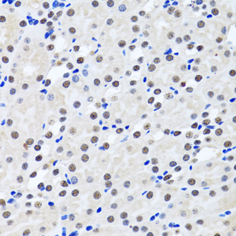
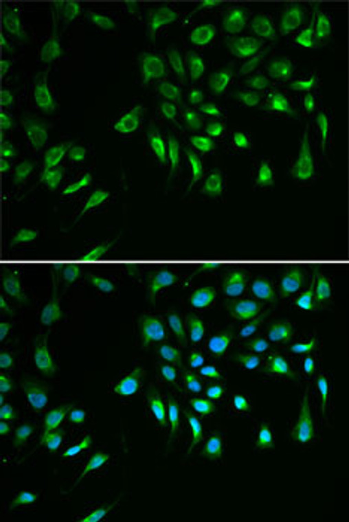
Rabbit polyclonal antibody anti-BIRC7 (1-265) is suitable for use in Western Blot, Immunohistochemistry and Immunofluorescence research applications. |
| Clonality: |
Polyclonal |
| Conjugation: |
Unconjugated |
| Isotype: |
IgG |
| Formulation: |
PBS with 0.02% Sodium Azide, 50% Glycerol, pH7.3. |
| Purification: |
Affinity purification |
| Dilution Range: |
WB 1:500-1:2000IHC-P 1:50-1:200IF/ICC 1:50-1:100 |
| Storage Instruction: |
Store at-20°C for up to 1 year from the date of receipt, and avoid repeat freeze-thaw cycles. |
| Gene Symbol: |
BIRC7 |
| Gene ID: |
79444 |
| Uniprot ID: |
BIRC7_HUMAN |
| Immunogen Region: |
1-265 |
| Immunogen: |
Recombinant fusion protein containing a sequence corresponding to amino acids 1-265 of human BIRC7 (NP_647478.1). |
| Immunogen Sequence: |
MGPKDSAKCLHRGPQPSHWA AGDGPTQERCGPRSLGSPVL GLDTCRAWDHVDGQILGQLR PLTEEEEEEGAGATLSRGPA FPGMGSEELRLASFYDWPLT AEVPPELLAAAGFFHTGHQD KVRCFFCYGGLQSWKRGDDP WTEHAKWFPSCQFLLRSKGR DFVHSVQETHSQLLGSWDPW EEPEDAAPVAPSVPASGYPE LPTPRREVQSESAQEPGGVS PAEAQRAWWVLEPPGARDV |
| Tissue Specificity | Isoform 1 and isoform 2 are expressed at very low levels or not detectable in most adult tissues. Detected in adult heart, placenta, lung, lymph node, spleen and ovary, and in several carcinoma cell lines. Isoform 2 is detected in fetal kidney, heart and spleen, and at lower levels in adult brain, skeletal muscle and peripheral blood leukocytes. |
| Post Translational Modifications | Autoubiquitinated and undergoes proteasome-mediated degradation. The truncated protein (tLivin) not only loses its anti-apoptotic effect but also acquires a pro-apoptotic effect. |
| Function | Apoptotic regulator capable of exerting proapoptotic and anti-apoptotic activities and plays crucial roles in apoptosis, cell proliferation, and cell cycle control. Its anti-apoptotic activity is mediated through the inhibition of CASP3, CASP7 and CASP9, as well as by its E3 ubiquitin-protein ligase activity. As it is a weak caspase inhibitor, its anti-apoptotic activity is thought to be due to its ability to ubiquitinate DIABLO/SMAC targeting it for degradation thereby promoting cell survival. May contribute to caspase inhibition, by blocking the ability of DIABLO/SMAC to disrupt XIAP/BIRC4-caspase interactions. Protects against apoptosis induced by TNF or by chemical agents such as adriamycin, etoposide or staurosporine. Suppression of apoptosis is mediated by activation of MAPK8/JNK1, and possibly also of MAPK9/JNK2. This activation depends on TAB1 and MAP3K7/TAK1. In vitro, inhibits CASP3 and proteolytic activation of pro-CASP9. Isoform 1: Blocks staurosporine-induced apoptosis. Promotes natural killer (NK) cell-mediated killing. Isoform 2: Blocks etoposide-induced apoptosis. Protects against natural killer (NK) cell-mediated killing. |
| Protein Name | Baculoviral Iap Repeat-Containing Protein 7Kidney Inhibitor Of Apoptosis ProteinKiapLivinMelanoma Inhibitor Of Apoptosis ProteinMl-IapRing Finger Protein 50Ring-Type E3 Ubiquitin Transferase Birc7 Cleaved Into - Baculoviral Iap Repeat-Containing Protein 7 30kda SubunitTruncated LivinP30-LivinTlivin |
| Cellular Localisation | NucleusCytoplasmGolgi ApparatusNuclearAnd In A Filamentous Pattern Throughout The CytoplasmFull-Length Livin Is Detected Exclusively In The CytoplasmWhereas The Truncated Form (Tlivin) Is Found In The Peri-Nuclear Region With Marked Localization To The Golgi ApparatusThe Accumulation Of Tlivin In The Nucleus Shows Positive Correlation With The Increase In Apoptosis |
| Alternative Antibody Names | Anti-Baculoviral Iap Repeat-Containing Protein 7 antibodyAnti-Kidney Inhibitor Of Apoptosis Protein antibodyAnti-Kiap antibodyAnti-Livin antibodyAnti-Melanoma Inhibitor Of Apoptosis Protein antibodyAnti-Ml-Iap antibodyAnti-Ring Finger Protein 50 antibodyAnti-Ring-Type E3 Ubiquitin Transferase Birc7 Cleaved Into - Baculoviral Iap Repeat-Containing Protein 7 30kda Subunit antibodyAnti-Truncated Livin antibodyAnti-P30-Livin antibodyAnti-Tlivin antibodyAnti-BIRC7 antibodyAnti-KIAP antibodyAnti-LIVIN antibodyAnti-MLIAP antibodyAnti-RNF50 antibodyAnti-UNQ5800 antibodyAnti-PRO19607 antibodyAnti-PRO21344 antibody |
Information sourced from Uniprot.org
12 months for antibodies. 6 months for ELISA Kits. Please see website T&Cs for further guidance


