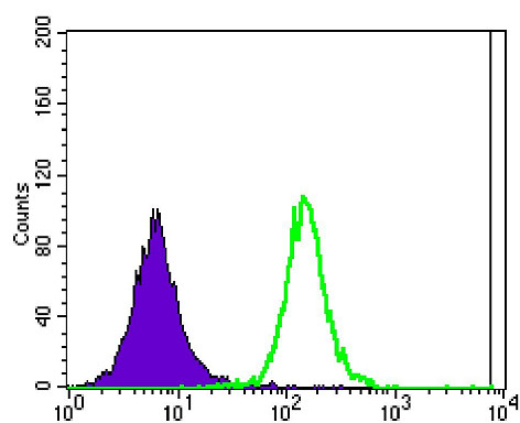
• Flow cytometric analysis of Jurkat cells using Ataxin-1 monoclonal antibody (green) and negative control (purple).
  • Immunohistochemistry analysis of paraffin-embedded ovarian cancer tissues (left) and lung cancer tissues (right) with DAB staining using Ataxin-1 monoclonal antibody.
  • Immunofluorescence analysis of NTERA-2 cells using Ataxin-1 monoclonal antibody (green). Blue: DRAQ5 fluorescent DNA dye. Red: Actin filaments have been labeled with Alexa Fluor-555 phalloidin.
  • Western blot analysis using Ataxin-1 monoclonal antibody against HEK293 (1) and ATXN1-hIgGFc transfected HEK293 (2) cell lysate.

Anti-ATXN1 antibody [2F5] (STJ97853)

SKU:
STJ97853

Current Stock:
Host: Mouse
Applications: WB/IHC/IF/FC/ELISA
Reactivity: Human
Note: STRICTLY FOR FURTHER SCIENTIFIC RESEARCH USE ONLY (RUO). MUST NOT TO BE USED IN DIAGNOSTIC OR THERAPEUTIC APPLICATIONS.
Short Description: Mouse monoclonal antibody anti-Ataxin-1 is suitable for use in Western Blot, Immunohistochemistry, Immunofluorescence, Flow Cytometry and ELISA research applications.
Clonality: Monoclonal
Clone ID: 2F5
Conjugation: Unconjugated
Isotype: IgG1
Formulation: Liquid in PBS containing 0.03% Sodium Azide, 0.5% BSA, 50% Glycerol.
Purification: Affinity purification
Dilution Range: WB 1:500-1:2000
IHC 1:200-1:1000
IF 1:200-1:1000
FC 1:200-1:400
ELISA 1:10000
Storage Instruction: Store at-20°C for up to 1 year from the date of receipt, and avoid repeat freeze-thaw cycles.
Gene Symbol: ATXN1
Gene ID: 6310
Uniprot ID: ATX1_HUMAN
Specificity: Ataxin-1 Monoclonal Antibody detects endogenous levels of Ataxin-1 protein.
Immunogen: Purified recombinant fragment of human Ataxin-1 expressed in E. Coli.
Post Translational Modifications Ubiquitinated by UBE3A, leading to its degradation by the proteasome. The presence of expanded poly-Gln repeats in spinocerebellar ataxia 1 (SCA1) patients impairs ubiquitination and degradation, leading to accumulation of ATXN1 in neurons and subsequent toxicity. Phosphorylation at Ser-775 increases the pathogenicity of proteins with an expanded polyglutamine tract. Sumoylation is dependent on nuclear localization and phosphorylation at Ser-775. It is reduced in the presence of an expanded polyglutamine tract.
Function Chromatin-binding factor that repress Notch signaling in the absence of Notch intracellular domain by acting as a CBF1 corepressor. Binds to the HEY promoter and might assist, along with NCOR2, RBPJ-mediated repression. Binds RNA in vitro. May be involved in RNA metabolism. In concert with CIC and ATXN1L, involved in brain development.
Protein Name Ataxin-1
Spinocerebellar Ataxia Type 1 Protein
Cellular Localisation Cytoplasm
Nucleus
Colocalizes With Usp7 In The Nucleus
Alternative Antibody Names Anti-Ataxin-1 antibody
Anti-Spinocerebellar Ataxia Type 1 Protein antibody
Anti-ATXN1 antibody
Anti-ATX1 antibody
Anti-SCA1 antibody

Information sourced from Uniprot.org

12 months for antibodies. 6 months for ELISA Kits. Please see website T&Cs for further guidance