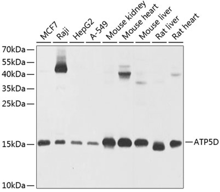
| Host: |
Rabbit |
| Applications: |
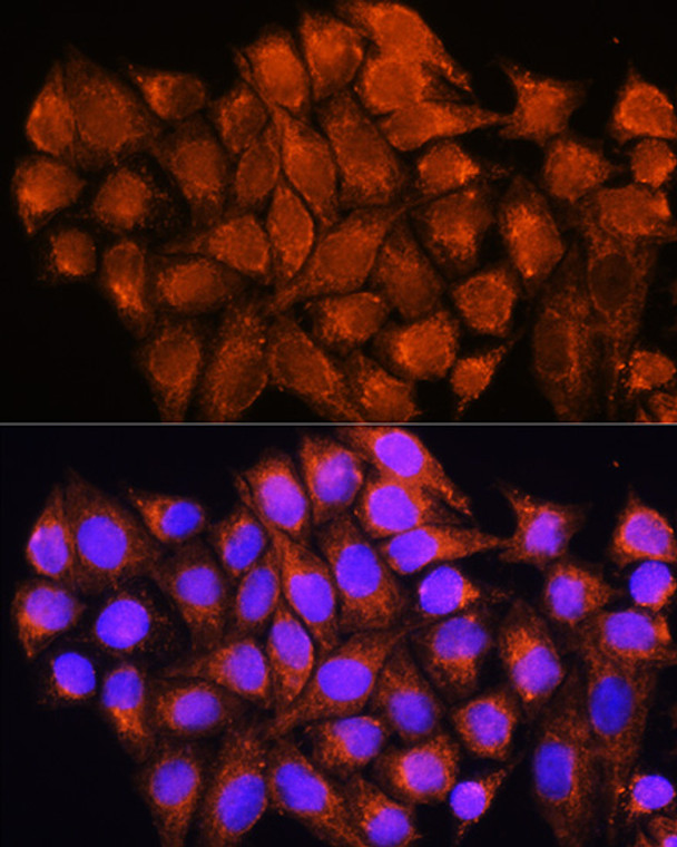
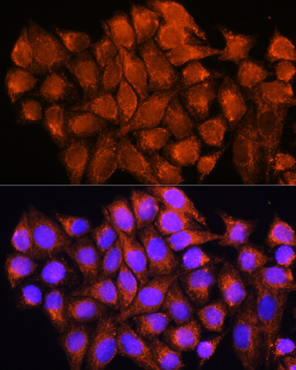
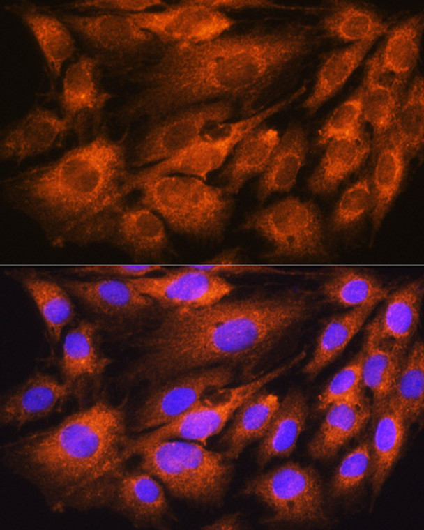
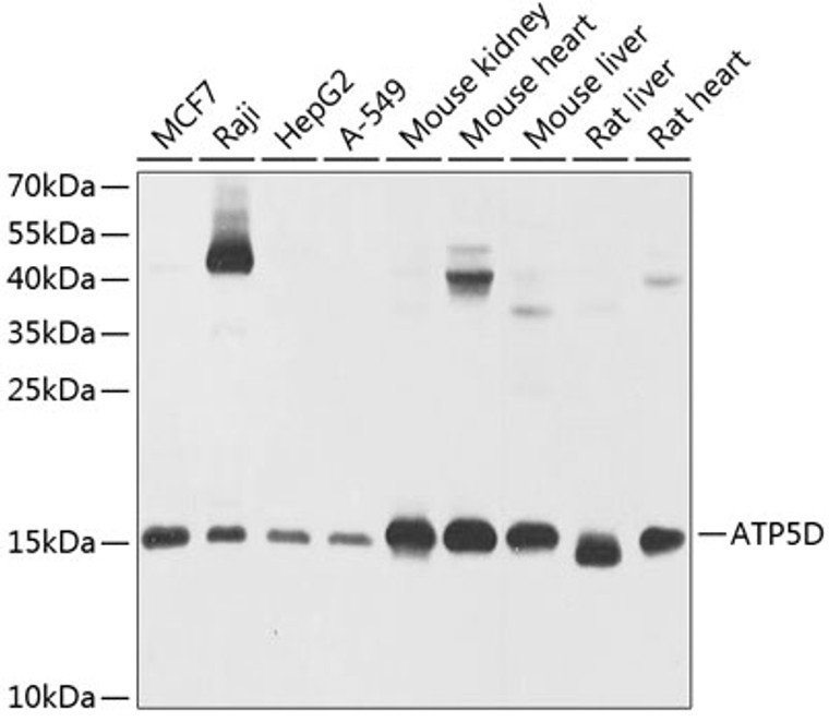
WB/IF |
| Reactivity: |
Human/Mouse/Rat |
| Note: |
STRICTLY FOR FURTHER SCIENTIFIC RESEARCH USE ONLY (RUO). MUST NOT TO BE USED IN DIAGNOSTIC OR THERAPEUTIC APPLICATIONS. |
| Short Description: |
Rabbit polyclonal antibody anti-ATP5D (1-168) is suitable for use in Western Blot and Immunofluorescence research applications. |
| Clonality: |
Polyclonal |
| Conjugation: |
Unconjugated |
| Isotype: |
IgG |
| Formulation: |
PBS with 0.02% Sodium Azide, 50% Glycerol, pH7.3. |
| Purification: |
Affinity purification |
| Dilution Range: |
WB 1:500-1:2000IF/ICC 1:50-1:200 |
| Storage Instruction: |
Store at-20°C for up to 1 year from the date of receipt, and avoid repeat freeze-thaw cycles. |
| Gene Symbol: |
ATP5F1D |
| Gene ID: |
513 |
| Uniprot ID: |
ATPD_HUMAN |
| Immunogen Region: |
1-168 |
| Immunogen: |
Recombinant fusion protein containing a sequence corresponding to amino acids 1-168 of human ATP5D (NP_001678.1). |
| Immunogen Sequence: |
MLPAALLRRPGLGRLVRHAR AYAEAAAAPAAASGPNQMSF TFASPTQVFFNGANVRQVDV PTLTGAFGILAAHVPTLQVL RPGLVVVHAEDGTTSKYFVS SGSIAVNADSSVQLLAEEAV TLDMLDLGAAKANLEKAQAE LVGTADEATRAEIQIRIEAN EALVKALE |
| Function | Mitochondrial membrane ATP synthase (F(1)F(0) ATP synthase or Complex V) produces ATP from ADP in the presence of a proton gradient across the membrane which is generated by electron transport complexes of the respiratory chain. F-type ATPases consist of two structural domains, F(1) - containing the extramembraneous catalytic core, and F(0) - containing the membrane proton channel, linked together by a central stalk and a peripheral stalk. During catalysis, ATP turnover in the catalytic domain of F(1) is coupled via a rotary mechanism of the central stalk subunits to proton translocation. Part of the complex F(1) domain and of the central stalk which is part of the complex rotary element. Rotation of the central stalk against the surrounding alpha(3)beta(3) subunits leads to hydrolysis of ATP in three separate catalytic sites on the beta subunits. |
| Protein Name | Atp Synthase Subunit Delta - MitochondrialAtp Synthase F1 Subunit DeltaF-Atpase Delta Subunit |
| Database Links | Reactome: R-HSA-163210Reactome: R-HSA-8949613 |
| Cellular Localisation | MitochondrionMitochondrion Inner Membrane |
| Alternative Antibody Names | Anti-Atp Synthase Subunit Delta - Mitochondrial antibodyAnti-Atp Synthase F1 Subunit Delta antibodyAnti-F-Atpase Delta Subunit antibodyAnti-ATP5F1D antibodyAnti-ATP5D antibody |
Information sourced from Uniprot.org
12 months for antibodies. 6 months for ELISA Kits. Please see website T&Cs for further guidance


