-
Immunohistochemical analysis of paraffin-embedded Mouse-kidney tissue. 1, AR Polyclonal Antibody was diluted at 1:200 (4°C, overnight). 2, Sodium citrate pH 6.0 was used for antibody retrieval (>98°C, 20min). 3, Secondary antibody was diluted at 1:200 (room tempeRature, 30min). Negative control was used by secondary antibody only.
-
Western blot analysis of Hela cells using AR Polyclonal Antibody.. Secondary antibody was diluted at 1:20000
-
Immunohistochemical analysis of paraffin-embedded Rat-heart tissue. 1, AR Polyclonal Antibody was diluted at 1:200 (4°C, overnight). 2, Sodium citrate pH 6.0 was used for antibody retrieval (>98°C, 20min). 3, Secondary antibody was diluted at 1:200 (room tempeRature, 30min). Negative control was used by secondary antibody only.
-
Immunohistochemical analysis of paraffin-embedded Mouse-liver tissue. 1, AR Polyclonal Antibody was diluted at 1:200 (4°C, overnight). 2, Sodium citrate pH 6.0 was used for antibody retrieval (>98°C, 20min). 3, Secondary antibody was diluted at 1:200 (room tempeRature, 30min). Negative control was used by secondary antibody only.
-
Western blot analysis of various cells using primary antibody diluted at 1:1000 (4°C overnight). Secondary antibody:Goat Anti-rabbit IgG IRDye 800 ( diluted at 1:5000, 25°C, 1 hour). Cell lysate was extracted by Minute Plasma Membrane Protein Isolation and Cell Fractionation Kit (SM-005, Inventbiotech, MN, USA).
-
Immunohistochemical analysis of paraffin-embedded Human-uterus tissue. 1, AR Polyclonal Antibody was diluted at 1:200 (4°C, overnight). 2, Sodium citrate pH 6.0 was used for antibody retrieval (>98°C, 20min). 3, Secondary antibody was diluted at 1:200 (room tempeRature, 30min). Negative control was used by secondary antibody only.
-
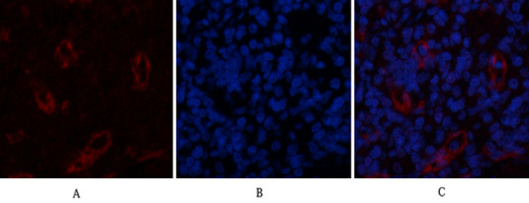
Immunofluorescence analysis of rat-spleen tissue. 1, AR Polyclonal Antibody (red) was diluted at 1:200 (4°C, overnight). 2, Cy3 labled Secondary antibody was diluted at 1:300 (room temperature, 50min).3, Picture B: DAPI (blue) 10min. Picture A:Target. Picture B: DAPI. Picture C: merge of A+B
-
Immunofluorescence analysis of rat-spleen tissue. 1, AR Polyclonal Antibody (red) was diluted at 1:200 (4°C, overnight). 2, Cy3 labled Secondary antibody was diluted at 1:300 (room temperature, 50min).3, Picture B: DAPI (blue) 10min. Picture A:Target. Picture B: DAPI. Picture C: merge of A+B
-
Immunofluorescence analysis of rat-heart tissue. 1, AR Polyclonal Antibody (red) was diluted at 1:200 (4°C, overnight). 2, Cy3 labled Secondary antibody was diluted at 1:300 (room temperature, 50min).3, Picture B: DAPI (blue) 10min. Picture A:Target. Picture B: DAPI. Picture C: merge of A+B
-
Immunofluorescence analysis of rat-heart tissue. 1, AR Polyclonal Antibody (red) was diluted at 1:200 (4°C, overnight). 2, Cy3 labled Secondary antibody was diluted at 1:300 (room temperature, 50min).3, Picture B: DAPI (blue) 10min. Picture A:Target. Picture B: DAPI. Picture C: merge of A+B
-
Immunofluorescence analysis of human-stomach tissue. 1, AR Polyclonal Antibody (red) was diluted at 1:200 (4°C, overnight). 2, Cy3 labled Secondary antibody was diluted at 1:300 (room temperature, 50min).3, Picture B: DAPI (blue) 10min. Picture A:Target. Picture B: DAPI. Picture C: merge of A+B
-
Immunofluorescence analysis of human-stomach tissue. 1, AR Polyclonal Antibody (red) was diluted at 1:200 (4°C, overnight). 2, Cy3 labled Secondary antibody was diluted at 1:300 (room temperature, 50min).3, Picture B: DAPI (blue) 10min. Picture A:Target. Picture B: DAPI. Picture C: merge of A+B
-
Immunofluorescence analysis of Hela cell. 1, AR Polyclonal Antibody (red) was diluted at 1:200 (4°C overnight). GFAP monoclonal antibody (5C8) (green) was diluted at 1:200 (4°C overnight). 2, Goat Anti Rabbit Alexa Fluor 594 Catalog: (NA was diluted at 1:1000 (room temperature, 50min). Goat Anti Mouse Alexa Fluor 488 Catalog: (NA was diluted at 1:1000 (room temperature, 50min).


![Anti-AR antibody (1-100) [S0MR] (STJ11101730) Anti-AR antibody (1-100) [S0MR] (STJ11101730)](https://cdn11.bigcommerce.com/s-zso2xnchw9/images/stencil/300x300/products/90645/360579/STJ11101730_1__89861.1713125258.png?c=1)


