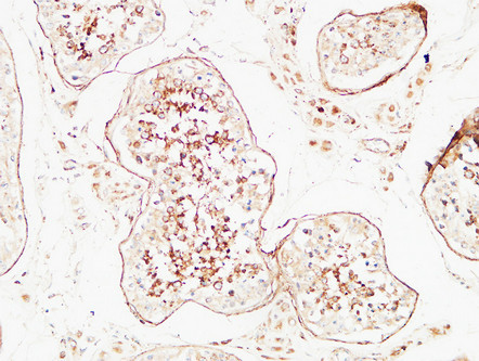
• Immunohistochemical analysis of paraffin-embedded Human testis. 1, Antibody was diluted at 1:200 (4°C overnight). 2, High-pressure and temperature EDTA, pH8.0 was used for antigen retrieval. 3, Secondary antibody was diluted at 1:200 (room temperature, 30min).
  • Immunohistochemical analysis of paraffin-embedded Human pancreas. 1, Antibody was diluted at 1:200 (4°C overnight). 2, High-pressure and temperature EDTA, pH8.0 was used for antigen retrieval. 3, Secondary antibody was diluted at 1:200 (room temperature, 30min).
  • Immunohistochemical analysis of paraffin-embedded Human testis. 1, Antibody was diluted at 1:200 (4°C overnight). 2, High-pressure and temperature EDTA, pH8.0 was used for antigen retrieval. 3, Secondary antibody was diluted at 1:200 (room temperature, 30min).
  • Immunohistochemical analysis of paraffin-embedded Human testis. 1, Antibody was diluted at 1:200 (4°C overnight). 2, High-pressure and temperature EDTA, pH8.0 was used for antigen retrieval. 3, Secondary antibody was diluted at 1:200 (room temperature, 30min).
  • Immunohistochemical analysis of paraffin-embedded human-lung-cancer, antibody was diluted at 1:200
  • Immunohistochemical analysis of paraffin-embedded human-lung-cancer, antibody was diluted at 1:200
  • Immunohistochemical analysis of paraffin-embedded human-colon-cancer, antibody was diluted at 1:200
  • Immunofluorescence analysis of human-heart tissue. 1, PTEN Polyclonal Antibody (red) was diluted at 1:200 (4°C, overnight). 2, Cy3 labled Secondary antibody was diluted at 1:300 (room temperature, 50min).3, Picture B: DAPI (blue) 10min. Picture A:Target. Picture B: DAPI. Picture C: merge of A+B
  • Immunofluorescence analysis of rat-spleen tissue. 1, PTEN Polyclonal Antibody (red) was diluted at 1:200 (4°C, overnight). 2, Cy3 labled Secondary antibody was diluted at 1:300 (room temperature, 50min).3, Picture B: DAPI (blue) 10min. Picture A:Target. Picture B: DAPI. Picture C: merge of A+B
  • Western blot analysis of mouse-heart mouse-brain mouse-lung Hela KB SH-SY5Y cells using PTEN Polyclonal Antibody diluted at 1:1000. Secondary antibody was diluted at 1:20000
  • Immunofluorescence analysis of rat-spleen tissue. 1, PTEN Polyclonal Antibody (red) was diluted at 1:200 (4°C, overnight). 2, Cy3 labled Secondary antibody was diluted at 1:300 (room temperature, 50min).3, Picture B: DAPI (blue) 10min. Picture A:Target. Picture B: DAPI. Picture C: merge of A+B
  • Immunofluorescence analysis of human-lung tissue. 1, PTEN Polyclonal Antibody (red) was diluted at 1:200 (4°C, overnight). 2, Cy3 labled Secondary antibody was diluted at 1:300 (room temperature, 50min).3, Picture B: DAPI (blue) 10min. Picture A:Target. Picture B: DAPI. Picture C: merge of A+B
  • Immunofluorescence analysis of human-lung tissue. 1, PTEN Polyclonal Antibody (red) was diluted at 1:200 (4°C, overnight). 2, Cy3 labled Secondary antibody was diluted at 1:300 (room temperature, 50min).3, Picture B: DAPI (blue) 10min. Picture A:Target. Picture B: DAPI. Picture C: merge of A+B
  • Immunohistochemical analysis of paraffin-embedded Human pancreas. 1, Antibody was diluted at 1:200 (4°C overnight). 2, High-pressure and temperature EDTA, pH8.0 was used for antigen retrieval. 3, Secondary antibody was diluted at 1:200 (room temperature, 30min).
  • Immunohistochemical analysis of paraffin-embedded human-colon-cancer, antibody was diluted at 1:200
  • Immunohistochemical analysis of paraffin-embedded Human pancreas. 1, Antibody was diluted at 1:200 (4°C overnight). 2, High-pressure and temperature EDTA, pH8.0 was used for antigen retrieval. 3, Secondary antibody was diluted at 1:200 (room temperature, 30min).
  • Immunofluorescence analysis of human-heart tissue. 1, PTEN Polyclonal Antibody (red) was diluted at 1:200 (4°C, overnight). 2, Cy3 labled Secondary antibody was diluted at 1:300 (room temperature, 50min).3, Picture B: DAPI (blue) 10min. Picture A:Target. Picture B: DAPI. Picture C: merge of A+B

Anti-PTEN antibody (251-300 aa) (STJ98583)

SKU:
STJ98583

Current Stock:
Host: Rabbit
Applications: IF/WB/IHC/ELISA
Reactivity: Human/Mouse/Rat
Note: STRICTLY FOR FURTHER SCIENTIFIC RESEARCH USE ONLY (RUO). MUST NOT TO BE USED IN DIAGNOSTIC OR THERAPEUTIC APPLICATIONS.
Short Description: Rabbit polyclonal antibody anti-Phosphatidylinositol 3, 4, 5-trisphosphate 3-phosphatase and dual-specificity protein phosphatase PTEN (251-300 aa) is suitable for use in Immunofluorescence, Western Blot, Immunohistochemistry and ELISA research appli
Clonality: Polyclonal
Conjugation: Unconjugated
Isotype: IgG
Formulation: Liquid in PBS containing 50% Glycerol, 0.5% BSA and 0.02% Sodium Azide.
Purification: The antibody was affinity-purified from rabbit antiserum by affinity-chromatography using epitope-specific immunogen.
Concentration: 1 mg/mL
Dilution Range: IF 1:50-200
IHC-P 100-300
WB 1:500-2000
ELISA 1:10000-20000
Storage Instruction: Store at-20°C for up to 1 year from the date of receipt, and avoid repeat freeze-thaw cycles.
Gene Symbol: PTEN
Gene ID: 5728
Uniprot ID: PTEN_HUMAN
Immunogen Region: 251-300 aa
Specificity: PTEN Polyclonal Antibody detects endogenous levels of PTEN
Immunogen: Synthesized peptide derived from PTEN at the amino acid range 251-300
Post Translational Modifications Constitutively phosphorylated by CK2 under normal conditions. Phosphorylated in vitro by MAST1, MAST2, MAST3 and STK11. Phosphorylation results in an inhibited activity towards PIP3. Phosphorylation can both inhibit or promote PDZ-binding. Phosphorylation at Tyr-336 by FRK/PTK5 protects this protein from ubiquitin-mediated degradation probably by inhibiting its binding to NEDD4. Phosphorylation by ROCK1 is essential for its stability and activity. Phosphorylation by PLK3 promotes its stability and prevents its degradation by the proteasome. Phosphorylated on Thr-319 and Thr-321 in the C2-type tensin domain following EGF stimulation which changes its binding preference from the p85 regulatory subunit of the PI3K kinase complex to DLC1. Monoubiquitinated.monoubiquitination is increased in presence of retinoic acid. Deubiquitinated by USP7.leading to its nuclear exclusion. Monoubiquitination of one of either Lys-13 and Lys-289 amino acid is sufficient to modulate PTEN compartmentalization. Ubiquitinated by XIAP/BIRC4. Ubiquitinated by the DCX(DCAF13) E3 ubiquitin ligase complex, leading to its degradation. ISGylated. ISGylation promotes PTEN degradation.
Function Acts as a dual-specificity protein phosphatase, dephosphorylating tyrosine-, serine- and threonine-phosphorylated proteins. Also acts as a lipid phosphatase, removing the phosphate in the D3 position of the inositol ring from phosphatidylinositol 3,4,5-trisphosphate, phosphatidylinositol 3,4-diphosphate, phosphatidylinositol 3-phosphate and inositol 1,3,4,5-tetrakisphosphate with order of substrate preference in vitro PtdIns(3,4,5)P3 > PtdIns(3,4)P2 > PtdIns3P > Ins(1,3,4,5)P4. Tumor suppressor, the lipid phosphatase activity is critical for its tumor suppressor function. Antagonizes the PI3K-AKT/PKB signaling pathway by dephosphorylating phosphoinositides and thereby modulating cell cycle progression and cell survival. The unphosphorylated form cooperates with MAGI2 to suppress AKT1 activation. In motile cells, suppresses the formation of lateral pseudopods and thereby promotes cell polarization and directed movement. Dephosphorylates tyrosine-phosphorylated focal adhesion kinase and inhibits cell migration and integrin-mediated cell spreading and focal adhesion formation. Required for growth factor-induced epithelial cell migration.growth factor stimulation induces PTEN phosphorylation which changes its binding preference from the p85 regulatory subunit of the PI3K kinase complex to DLC1 and results in translocation of the PTEN-DLC1 complex to the posterior of migrating cells to promote RHOA activation. Meanwhile, TNS3 switches binding preference from DLC1 to p85 and the TNS3-p85 complex translocates to the leading edge of migrating cells to activate RAC1 activation. Plays a role as a key modulator of the AKT-mTOR signaling pathway controlling the tempo of the process of newborn neurons integration during adult neurogenesis, including correct neuron positioning, dendritic development and synapse formation. Involved in the regulation of synaptic function in excitatory hippocampal synapses. Recruited to the postsynaptic membrane upon NMDA receptor activation, is required for the modulation of synaptic activity during plasticity. Enhancement of lipid phosphatase activity is able to drive depression of AMPA receptor-mediated synaptic responses, activity required for NMDA receptor-dependent long-term depression (LTD). May be a negative regulator of insulin signaling and glucose metabolism in adipose tissue. The nuclear monoubiquitinated form possesses greater apoptotic potential, whereas the cytoplasmic nonubiquitinated form induces less tumor suppressive ability. Isoform alpha: Functional kinase, like isoform 1 it antagonizes the PI3K-AKT/PKB signaling pathway. Plays a role in mitochondrial energetic metabolism by promoting COX activity and ATP production, via collaboration with isoform 1 in increasing protein levels of PINK1.
Protein Name Phosphatidylinositol 3 -4 -5-Trisphosphate 3-Phosphatase And Dual-Specificity Protein Phosphatase Pten
Mutated In Multiple Advanced Cancers 1
Phosphatase And Tensin Homolog
Database Links Reactome: R-HSA-1660499
Reactome: R-HSA-1855204
Reactome: R-HSA-199418
Reactome: R-HSA-202424
Reactome: R-HSA-5628897
Reactome: R-HSA-5674404
Reactome: R-HSA-5689880
Reactome: R-HSA-5689896
Reactome: R-HSA-8943723
Reactome: R-HSA-8948747
Reactome: R-HSA-8948751
Reactome: R-HSA-8986944
Cellular Localisation Cytoplasm
Nucleus
Pml Body
Cell Projection
Dendritic Spine
Postsynaptic Density
Monoubiquitinated Form Is Nuclear
Nonubiquitinated Form Is Cytoplasmic
Colocalized With Pml And Usp7 In Pml Nuclear Bodies
Xiap/Birc4 Promotes Its Nuclear Localization
Associares With The Postsynaptic Density In Response To Nmdar Activation
Isoform Alpha: Secreted
May Be Secreted Via A Classical Signal Peptide And Reenter Into Cells With The Help Of A Poly-Arg Motif
Alternative Antibody Names Anti-Phosphatidylinositol 3 -4 -5-Trisphosphate 3-Phosphatase And Dual-Specificity Protein Phosphatase Pten antibody
Anti-Mutated In Multiple Advanced Cancers 1 antibody
Anti-Phosphatase And Tensin Homolog antibody
Anti-PTEN antibody
Anti-MMAC1 antibody
Anti-TEP1 antibody

Information sourced from Uniprot.org

12 months for antibodies. 6 months for ELISA Kits. Please see website T&Cs for further guidance