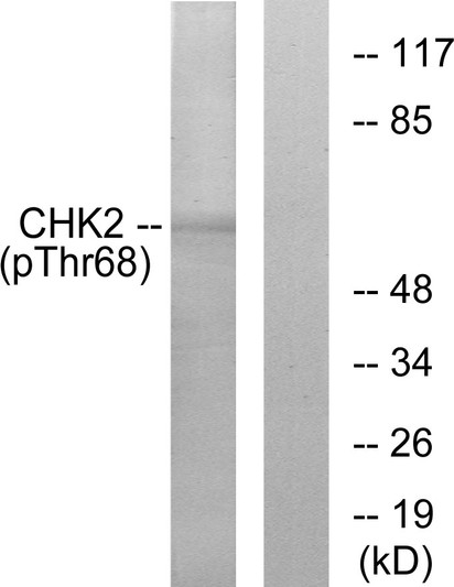
• Western blot analysis of lysates from Jurkat cells treated with UV, using Chk2 (Phospho-Thr68) Antibody. The lane on the right is blocked with the phospho peptide.
  • Western blot analysis of lysates from 1) 293T, 2) HELA cells, (Green) primary antibody was diluted at 1:1000, 4°C over night, secondary antibody (cat: (NA) was diluted at 1:10000, 37°C 1hour. (Red) Actin Beta monoclonal antibody (5B7) (cat: (STJ96930) antibody was diluted at 1:5000 as loading control, 4°C over night, secondary antibody (cat: (NA) was diluted at 1:10000, 37°C 1hour.
  • Western blot analysis of various lysis using Phospho-Chk2 (T68) Polyclonal Antibody diluted at 1:500. Secondary antibody was diluted at 1:20000 cells nucleus extracted by Minute TM Cytoplasmic and Nuclear Fractionation kit (SC-003, Inventbiotech, MN, USA).
  • Immunohistochemistry analysis of paraffin-embedded human lung carcinoma, using Chk2 (Phospho-Thr68) Antibody. The picture on the right is blocked with the phospho peptide.
  • Western blot analysis of various cells using Phospho-Chk2 (T68) Polyclonal Antibody diluted at 1:500 cells nucleus extracted by Minute TM Cytoplasmic and Nuclear Fractionation kit (SC-003, Inventbiotech, MN, USA).
  • Western blot analysis of HELA cells using Phospho-Chk2 (T68) Polyclonal Antibody diluted at 1:500 cells nucleus extracted by Minute TM Cytoplasmic and Nuclear Fractionation kit (SC-003, Inventbiotech, MN, USA).
  • Immunohistochemical analysis of paraffin-embedded Rat-lung tissue. 1, Chk2 (phospho Thr68) Polyclonal Antibody was diluted at 1:200 (4°C, overnight). 2, Sodium citrate pH 6.0 was used for antibody retrieval (>98°C, 20min). 3, Secondary antibody was diluted at 1:200 (room tempeRature, 30min). Negative control was used by secondary antibody only.
  • Immunohistochemical analysis of paraffin-embedded Mouse-lung tissue. 1, Chk2 (phospho Thr68) Polyclonal Antibody was diluted at 1:200 (4°C, overnight). 2, Sodium citrate pH 6.0 was used for antibody retrieval (>98°C, 20min). 3, Secondary antibody was diluted at 1:200 (room tempeRature, 30min). Negative control was used by secondary antibody only.
  • Immunohistochemical analysis of paraffin-embedded Human-lung tissue. 1, Chk2 (phospho Thr68) Polyclonal Antibody was diluted at 1:200 (4°C, overnight). 2, Sodium citrate pH 6.0 was used for antibody retrieval (>98°C, 20min). 3, Secondary antibody was diluted at 1:200 (room tempeRature, 30min). Negative control was used by secondary antibody only.
  • Immunohistochemical analysis of paraffin-embedded Human-uterus tissue. 1, Chk2 (phospho Thr68) Polyclonal Antibody was diluted at 1:200 (4°C, overnight). 2, Sodium citrate pH 6.0 was used for antibody retrieval (>98°C, 20min). 3, Secondary antibody was diluted at 1:200 (room tempeRature, 30min). Negative control was used by secondary antibody only.
  • Immunofluorescence analysis of rat-lung tissue. 1, Chk2 (phospho Thr68) Polyclonal Antibody (red) was diluted at 1:200 (4°C, overnight). 2, Cy3 labled Secondary antibody was diluted at 1:300 (room temperature, 50min).3, Picture B: DAPI (blue) 10min. Picture A:Target. Picture B: DAPI. Picture C: merge of A+B
  • Immunofluorescence analysis of rat-heart tissue. 1, Chk2 (phospho Thr68) Polyclonal Antibody (red) was diluted at 1:200 (4°C, overnight). 2, Cy3 labled Secondary antibody was diluted at 1:300 (room temperature, 50min).3, Picture B: DAPI (blue) 10min. Picture A:Target. Picture B: DAPI. Picture C: merge of A+B
  • Immunofluorescence analysis of Hela cell. 1, Chk2 (phospho Thr68) Polyclonal Antibody (red) was diluted at 1:200 (4°C overnight). HAO1 monoclonal antibody (Mix) (green) was diluted at 1:200 (4°C overnight). 2, Goat Anti Rabbit Alexa Fluor 594 Catalog: (NA was diluted at 1:1000 (room temperature, 50min). Goat Anti Mouse Alexa Fluor 488 Catalog: (NA was diluted at 1:1000 (room temperature, 50min).

Anti-Phospho-CHEK2-Thr68 antibody (35-84 aa) (STJ90225)

SKU:
STJ90225

Current Stock:
Host: Rabbit
Applications: IF/WB/IHC/ELISA
Reactivity: Human/Mouse/Rat
Note: STRICTLY FOR FURTHER SCIENTIFIC RESEARCH USE ONLY (RUO). MUST NOT TO BE USED IN DIAGNOSTIC OR THERAPEUTIC APPLICATIONS.
Short Description: Rabbit polyclonal antibody anti-Phospho-Serine/threonine-protein kinase Chk2-Thr68 (35-84 aa) is suitable for use in Immunofluorescence, Western Blot, Immunohistochemistry and ELISA research applications.
Clonality: Polyclonal
Conjugation: Unconjugated
Isotype: IgG
Formulation: Liquid in PBS containing 50% Glycerol, 0.5% BSA and 0.02% Sodium Azide.
Purification: The antibody was affinity-purified from rabbit antiserum by affinity-chromatography using epitope-specific immunogen.
Concentration: 1 mg/mL
Dilution Range: IF 1:50-200
WB 1:500-1:2000
IHC 1:100-1:300
ELISA 1:20000
Storage Instruction: Store at-20°C for up to 1 year from the date of receipt, and avoid repeat freeze-thaw cycles.
Gene Symbol: CHEK2
Gene ID: 11200
Uniprot ID: CHK2_HUMAN
Immunogen Region: 35-84 aa
Specificity: Phospho-Chk2 (T68) Polyclonal Antibody detects endogenous levels of Chk2 protein only when phosphorylated at T68.
Immunogen: The antiserum was produced against synthesized peptide derived from the human Chk2 around the phosphorylation site of Thr68 at the amino acid range 35-84
Post Translational Modifications Phosphorylated. Phosphorylated at Ser-73 by PLK3 in response to DNA damage, promoting phosphorylation at Thr-68 by ATM and the G2/M transition checkpoint. Phosphorylation at Thr-68 induces homodimerization. Autophosphorylates at Thr-383 and Thr-387 in the T-loop/activation segment upon dimerization to become fully active and phosphorylate its substrates like for instance CDC25C. DNA damage-induced autophosphorylation at Ser-379 induces CUL1-mediated ubiquitination and regulates the pro-apoptotic function. Phosphorylation at Ser-456 also regulates ubiquitination. Phosphorylated by PLK4. Ubiquitinated. CUL1-mediated ubiquitination regulates the pro-apoptotic function. Ubiquitination may also regulate protein stability. Ubiquitinated by RNF8 via 'Lys-48'-linked ubiquitination.
Function Serine/threonine-protein kinase which is required for checkpoint-mediated cell cycle arrest, activation of DNA repair and apoptosis in response to the presence of DNA double-strand breaks. May also negatively regulate cell cycle progression during unperturbed cell cycles. Following activation, phosphorylates numerous effectors preferentially at the consensus sequence L-X-R-X-X-S/T. Regulates cell cycle checkpoint arrest through phosphorylation of CDC25A, CDC25B and CDC25C, inhibiting their activity. Inhibition of CDC25 phosphatase activity leads to increased inhibitory tyrosine phosphorylation of CDK-cyclin complexes and blocks cell cycle progression. May also phosphorylate NEK6 which is involved in G2/M cell cycle arrest. Regulates DNA repair through phosphorylation of BRCA2, enhancing the association of RAD51 with chromatin which promotes DNA repair by homologous recombination. Also stimulates the transcription of genes involved in DNA repair (including BRCA2) through the phosphorylation and activation of the transcription factor FOXM1. Regulates apoptosis through the phosphorylation of p53/TP53, MDM4 and PML. Phosphorylation of p53/TP53 at 'Ser-20' by CHEK2 may alleviate inhibition by MDM2, leading to accumulation of active p53/TP53. Phosphorylation of MDM4 may also reduce degradation of p53/TP53. Also controls the transcription of pro-apoptotic genes through phosphorylation of the transcription factor E2F1. Tumor suppressor, it may also have a DNA damage-independent function in mitotic spindle assembly by phosphorylating BRCA1. Its absence may be a cause of the chromosomal instability observed in some cancer cells. Promotes the CCAR2-SIRT1 association and is required for CCAR2-mediated SIRT1 inhibition. (Microbial infection) Phosphorylates herpes simplex virus 1/HHV-1 protein ICP0 and thus activates its SUMO-targeted ubiquitin ligase activity.
Protein Name Serine/Threonine-Protein Kinase Chk2
Chk2 Checkpoint Homolog
Cds1 Homolog
Hucds1
Hcds1
Checkpoint Kinase 2
Database Links Reactome: R-HSA-5693565
Reactome: R-HSA-6804756
Reactome: R-HSA-6804757
Reactome: R-HSA-6804760
Reactome: R-HSA-69473
Reactome: R-HSA-69541
Reactome: R-HSA-69601
Reactome: R-HSA-75035
Cellular Localisation Isoform 2: Nucleus
Isoform 10 Is Present Throughout The Cell
Isoform 4: Nucleus
Isoform 7: Nucleus
Isoform 9: Nucleus
Isoform 12: Nucleus
Nucleus
Pml Body
Nucleoplasm
Recruited Into Pml Bodies Together With Tp53
Alternative Antibody Names Anti-Serine/Threonine-Protein Kinase Chk2 antibody
Anti-Chk2 Checkpoint Homolog antibody
Anti-Cds1 Homolog antibody
Anti-Hucds1 antibody
Anti-Hcds1 antibody
Anti-Checkpoint Kinase 2 antibody
Anti-CHEK2 antibody
Anti-CDS1 antibody
Anti-CHK2 antibody
Anti-RAD53 antibody

Information sourced from Uniprot.org

12 months for antibodies. 6 months for ELISA Kits. Please see website T&Cs for further guidance