• Immunohistochemical analysis of paraffin-embedded Human lymphoma. 1, Antibody was diluted at 1:200 (4°C overnight). 2, High-pressure and temperature EDTA, pH8.0 was used for antigen retrieval. 3, Secondary antibody was diluted at 1:200 (room temperature, 30min).
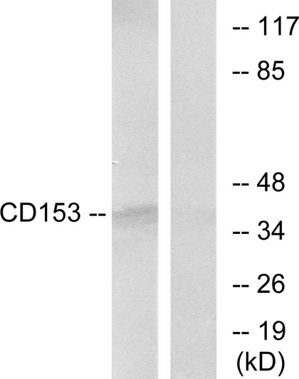
  • Western blot analysis of lysate from THP-1 cells, using IL10 Antibody.
  • Immunohistochemical analysis of paraffin-embedded Human lymphoma. 1, Antibody was diluted at 1:200 (4°C overnight). 2, High-pressure and temperature EDTA, pH8.0 was used for antigen retrieval. 3, Secondary antibody was diluted at 1:200 (room temperature, 30min).
  • Immunohistochemical analysis of paraffin-embedded Human lymphoma. 1, Antibody was diluted at 1:200 (4°C overnight). 2, High-pressure and temperature EDTA, pH8.0 was used for antigen retrieval. 3, Secondary antibody was diluted at 1:200 (room temperature, 30min).
  • Immunohistochemical analysis of paraffin-embedded Human Cerebral cortex. 1, Antibody was diluted at 1:200 (4°C overnight). 2, High-pressure and temperature EDTA, pH8.0 was used for antigen retrieval. 3, Secondary antibody was diluted at 1:200 (room temperature, 30min).
  • Immunohistochemical analysis of paraffin-embedded Human Cerebral cortex. 1, Antibody was diluted at 1:200 (4°C overnight). 2, High-pressure and temperature EDTA, pH8.0 was used for antigen retrieval. 3, Secondary antibody was diluted at 1:200 (room temperature, 30min).
  • Immunohistochemical analysis of paraffin-embedded Human Cerebral cortex. 1, Antibody was diluted at 1:200 (4°C overnight). 2, High-pressure and temperature EDTA, pH8.0 was used for antigen retrieval. 3, Secondary antibody was diluted at 1:200 (room temperature, 30min).
  • Immunohistochemical analysis of paraffin-embedded human-breast-cancer, antibody was diluted at 1:100
  • Western blot analysis of THP-1, Romas, HepG2, SW480, SHSY5Y, Mouse liver cells using IL-10 Polyclonal Antibody.. Secondary antibody was diluted at 1:20000

Anti-IL10 antibody (71-120 aa) (STJ96503)

SKU:
STJ96503

Current Stock:
Host: Rabbit
Applications: WB/IHC/IF/ELISA
Reactivity: Human/Mouse/Rat
Note: STRICTLY FOR FURTHER SCIENTIFIC RESEARCH USE ONLY (RUO). MUST NOT TO BE USED IN DIAGNOSTIC OR THERAPEUTIC APPLICATIONS.
Short Description: Rabbit polyclonal antibody anti-Interleukin-10 (71-120 aa) is suitable for use in Western Blot, Immunohistochemistry, Immunofluorescence and ELISA research applications.
Clonality: Polyclonal
Conjugation: Unconjugated
Isotype: IgG
Formulation: Liquid in PBS containing 50% Glycerol, 0.5% BSA and 0.02% Sodium Azide.
Purification: The antibody was affinity-purified from rabbit antiserum by affinity-chromatography using epitope-specific immunogen.
Concentration: 1 mg/mL
Dilution Range: WB 1:500-1:2000
IHC-P 1:100-300
ELISA 1:20000
IF 1:100-300
Storage Instruction: Store at-20°C for up to 1 year from the date of receipt, and avoid repeat freeze-thaw cycles.
Gene Symbol: IL10
Gene ID: 3586
Uniprot ID: IL10_HUMAN
Immunogen Region: 71-120 aa
Specificity: IL-10 Polyclonal Antibody detects endogenous levels of IL-10 protein.
Immunogen: The antiserum was produced against synthesized peptide derived from the Internal region of human IL10 at the amino acid range 71-120
Function Major immune regulatory cytokine that acts on many cells of the immune system where it has profound anti-inflammatory functions, limiting excessive tissue disruption caused by inflammation. Mechanistically, IL10 binds to its heterotetrameric receptor comprising IL10RA and IL10RB leading to JAK1 and STAT2-mediated phosphorylation of STAT3. In turn, STAT3 translocates to the nucleus where it drives expression of anti-inflammatory mediators. Targets antigen-presenting cells (APCs) such as macrophages and monocytes and inhibits their release of pro-inflammatory cytokines including granulocyte-macrophage colony-stimulating factor /GM-CSF, granulocyte colony-stimulating factor/G-CSF, IL-1 alpha, IL-1 beta, IL-6, IL-8 and TNF-alpha. Interferes also with antigen presentation by reducing the expression of MHC-class II and co-stimulatory molecules, thereby inhibiting their ability to induce T cell activation. In addition, controls the inflammatory response of macrophages by reprogramming essential metabolic pathways including mTOR signaling.
Protein Name Interleukin-10
Il-10
Cytokine Synthesis Inhibitory Factor
Csif
Database Links Reactome: R-HSA-6783783
Reactome: R-HSA-6785807
Reactome: R-HSA-8950505
Reactome: R-HSA-9662834
Reactome: R-HSA-9664323
Cellular Localisation Secreted
Alternative Antibody Names Anti-Interleukin-10 antibody
Anti-Il-10 antibody
Anti-Cytokine Synthesis Inhibitory Factor antibody
Anti-Csif antibody
Anti-IL10 antibody

Information sourced from Uniprot.org

12 months for antibodies. 6 months for ELISA Kits. Please see website T&Cs for further guidance