• Immunofluorescence analysis of rat-lung tissue. 1, NF Kappa B-p65 (phospho Ser536) Polyclonal Antibody (red) was diluted at 1:200 (4°C, overnight). 2, Cy3 labled Secondary antibody was diluted at 1:300 (room temperature, 50min).3, Picture B: DAPI (blue) 10min. Picture A:Target. Picture B: DAPI. Picture C: merge of A+B
  • Immunohistochemical analysis of paraffin-embedded Rat-testis tissue. 1, NF Kappa B-p65 (phospho Ser536) Polyclonal Antibody was diluted at 1:200 (4°C, overnight). 2, Sodium citrate pH 6.0 was used for antibody retrieval (>98°C, 20min). 3, Secondary antibody was diluted at 1:200 (room tempeRature, 30min). Negative control was used by secondary antibody only.
  • Western blot analysis of A549 3T3 293T K562 cells using Antibody diluted at 2000. Secondary antibody was diluted at 1:20000
  • Western blot analysis of lysates from K562 cells and COLO cells, using NF-kappaB p65 (Phospho-Ser536) Antibody. The lane on the right is blocked with the phospho peptide.
  • Immunohistochemical analysis of paraffin-embedded Mouse-brain tissue. 1, NF Kappa B-p65 (phospho Ser536) Polyclonal Antibody was diluted at 1:200 (4°C, overnight). 2, Sodium citrate pH 6.0 was used for antibody retrieval (>98°C, 20min). 3, Secondary antibody was diluted at 1:200 (room tempeRature, 30min). Negative control was used by secondary antibody only.
  • Western blot analysis of various cells using Phospho-NF Kappa B-p65 (S536) Polyclonal Antibody diluted at 1:2000
  • Immunohistochemical analysis of paraffin-embedded Human-uterus tissue. 1, NF Kappa B-p65 (phospho Ser536) Polyclonal Antibody was diluted at 1:200 (4°C, overnight). 2, Sodium citrate pH 6.0 was used for antibody retrieval (>98°C, 20min). 3, Secondary antibody was diluted at 1:200 (room tempeRature, 30min). Negative control was used by secondary antibody only.
  • Immunohistochemical analysis of paraffin-embedded Mouse-liver tissue. 1, NF Kappa B-p65 (phospho Ser536) Polyclonal Antibody was diluted at 1:200 (4°C, overnight). 2, Sodium citrate pH 6.0 was used for antibody retrieval (>98°C, 20min). 3, Secondary antibody was diluted at 1:200 (room tempeRature, 30min). Negative control was used by secondary antibody only.
  • Immunofluorescence analysis of mouse-kidney tissue. 1, NF Kappa B-p65 (phospho Ser536) Polyclonal Antibody (red) was diluted at 1:200 (4°C, overnight). 2, Cy3 labled Secondary antibody was diluted at 1:300 (room temperature, 50min).3, Picture B: DAPI (blue) 10min. Picture A:Target. Picture B: DAPI. Picture C: merge of A+B
  • Immunofluorescence analysis of mouse-kidney tissue. 1, NF Kappa B-p65 (phospho Ser536) Polyclonal Antibody (red) was diluted at 1:200 (4°C, overnight). 2, Cy3 labled Secondary antibody was diluted at 1:300 (room temperature, 50min).3, Picture B: DAPI (blue) 10min. Picture A:Target. Picture B: DAPI. Picture C: merge of A+B
  • Immunofluorescence analysis of rat-spleen tissue. 1, NF Kappa B-p65 (phospho Ser536) Polyclonal Antibody (red) was diluted at 1:200 (4°C, overnight). 2, Cy3 labled Secondary antibody was diluted at 1:300 (room temperature, 50min).3, Picture B: DAPI (blue) 10min. Picture A:Target. Picture B: DAPI. Picture C: merge of A+B
  • Immunofluorescence analysis of rat-spleen tissue. 1, NF Kappa B-p65 (phospho Ser536) Polyclonal Antibody (red) was diluted at 1:200 (4°C, overnight). 2, Cy3 labled Secondary antibody was diluted at 1:300 (room temperature, 50min).3, Picture B: DAPI (blue) 10min. Picture A:Target. Picture B: DAPI. Picture C: merge of A+B
  • Immunofluorescence analysis of rat-lung tissue. 1, NF Kappa B-p65 (phospho Ser536) Polyclonal Antibody (red) was diluted at 1:200 (4°C, overnight). 2, Cy3 labled Secondary antibody was diluted at 1:300 (room temperature, 50min).3, Picture B: DAPI (blue) 10min. Picture A:Target. Picture B: DAPI. Picture C: merge of A+B
  • Immunohistochemical analysis of paraffin-embedded Rat-lung tissue. 1, NF Kappa B-p65 (phospho Ser536) Polyclonal Antibody was diluted at 1:200 (4°C, overnight). 2, Sodium citrate pH 6.0 was used for antibody retrieval (>98°C, 20min). 3, Secondary antibody was diluted at 1:200 (room tempeRature, 30min). Negative control was used by secondary antibody only.
  • Immunohistochemical analysis of paraffin-embedded Human-uterus-cancer tissue. 1, NF Kappa B-p65 (phospho Ser536) Polyclonal Antibody was diluted at 1:200 (4°C, overnight). 2, Sodium citrate pH 6.0 was used for antibody retrieval (>98°C, 20min). 3, Secondary antibody was diluted at 1:200 (room tempeRature, 30min). Negative control was used by secondary antibody only.
  • Immunohistochemical analysis of paraffin-embedded Rat-spleen tissue. 1, NF Kappa B-p65 (phospho Ser536) Polyclonal Antibody was diluted at 1:200 (4°C, overnight). 2, Sodium citrate pH 6.0 was used for antibody retrieval (>98°C, 20min). 3, Secondary antibody was diluted at 1:200 (room tempeRature, 30min). Negative control was used by secondary antibody only.
  • Immunohistochemical analysis of paraffin-embedded Human-colon tissue. 1, NF Kappa B-p65 (phospho Ser536) Polyclonal Antibody was diluted at 1:200 (4°C, overnight). 2, Sodium citrate pH 6.0 was used for antibody retrieval (>98°C, 20min). 3, Secondary antibody was diluted at 1:200 (room tempeRature, 30min). Negative control was used by secondary antibody only.
  • Immunohistochemical analysis of paraffin-embedded Mouse-lung tissue. 1, NF Kappa B-p65 (phospho Ser536) Polyclonal Antibody was diluted at 1:200 (4°C, overnight). 2, Sodium citrate pH 6.0 was used for antibody retrieval (>98°C, 20min). 3, Secondary antibody was diluted at 1:200 (room tempeRature, 30min). Negative control was used by secondary antibody only.
  • Immunohistochemical analysis of paraffin-embedded Human-lung tissue. 1, NF Kappa B-p65 (phospho Ser536) Polyclonal Antibody was diluted at 1:200 (4°C, overnight). 2, Sodium citrate pH 6.0 was used for antibody retrieval (>98°C, 20min). 3, Secondary antibody was diluted at 1:200 (room tempeRature, 30min). Negative control was used by secondary antibody only.
  • Immunohistochemical analysis of paraffin-embedded Mouse-spleen tissue. 1, NF Kappa B-p65 (phospho Ser536) Polyclonal Antibody was diluted at 1:200 (4°C, overnight). 2, Sodium citrate pH 6.0 was used for antibody retrieval (>98°C, 20min). 3, Secondary antibody was diluted at 1:200 (room tempeRature, 30min). Negative control was used by secondary antibody only.
  • Immunohistochemical analysis of paraffin-embedded Human-lung-cancer tissue. 1, NF Kappa B-p65 (phospho Ser536) Polyclonal Antibody was diluted at 1:200 (4°C, overnight). 2, Sodium citrate pH 6.0 was used for antibody retrieval (>98°C, 20min). 3, Secondary antibody was diluted at 1:200 (room tempeRature, 30min). Negative control was used by secondary antibody only.
  • Western blot analysis of COLO205 cells using Phospho-NF Kappa B-p65 (S536) Polyclonal Antibody diluted at 1:2000
  • Immunohistochemical analysis of paraffin-embedded Human-stomach-cancer tissue. 1, NF Kappa B-p65 (phospho Ser536) Polyclonal Antibody was diluted at 1:200 (4°C, overnight). 2, Sodium citrate pH 6.0 was used for antibody retrieval (>98°C, 20min). 3, Secondary antibody was diluted at 1:200 (room tempeRature, 30min). Negative control was used by secondary antibody only.
  • Immunohistochemistry analysis of paraffin-embedded human breast carcinoma, using NF-kappaB p65 (Phospho-Ser536) Antibody. The picture on the right is blocked with the phospho peptide.
  • Immunohistochemical analysis of paraffin-embedded Human-Appendix tissue. 1, NF Kappa B-p65 (phospho Ser536) Polyclonal Antibody was diluted at 1:200 (4°C, overnight). 2, Sodium citrate pH 6.0 was used for antibody retrieval (>98°C, 20min). 3, Secondary antibody was diluted at 1:200 (room tempeRature, 30min). Negative control was used by secondary antibody only.
  • Western blot analysis of lysates from 1) A549, 2) 3T3, 3) 293T, 4) K562 cells, (Green) primary antibody was diluted at 1:1000, 4°C over night, secondary antibody (cat: (NA) was diluted at 1:10000, 37°C 1hour. (Red) Actin Beta monoclonal antibody (5B7) (cat: (STJ96930) antibody was diluted at 1:5000 as loading control, 4°C over night, secondary antibody (cat: (NA) was diluted at 1:10000, 37°C 1hour.
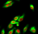
  • Immunofluorescence analysis of Hela cell. 1, NF Kappa B-p65 (phospho Ser536) Polyclonal Antibody (red) was diluted at 1:200 (4°C overnight). Alpha-tubulin monoclonal antibody (8F11) (green) was diluted at 1:200 (4°C overnight). 2, Goat Anti Rabbit Alexa Fluor 594 Catalog: (NA was diluted at 1:1000 (room temperature, 50min). Goat Anti Mouse Alexa Fluor 488 Catalog: (NA was diluted at 1:1000 (room temperature, 50min).

Anti-Phospho-RELA-Ser536 antibody (502-551 aa) (STJ90351)

SKU:
STJ90351

Current Stock:
Host: Rabbit
Applications: IF/WB/IHC/IP/ELISA
Reactivity: Human/Mouse/Rat/Monkey
Note: STRICTLY FOR FURTHER SCIENTIFIC RESEARCH USE ONLY (RUO). MUST NOT TO BE USED IN DIAGNOSTIC OR THERAPEUTIC APPLICATIONS.
Short Description: Rabbit polyclonal antibody anti-Phospho-Transcription factor p65-Ser536 (502-551 aa) is suitable for use in Immunofluorescence, Western Blot, Immunohistochemistry, Immunoprecipitation and ELISA research applications.
Clonality: Polyclonal
Conjugation: Unconjugated
Isotype: IgG
Formulation: Liquid in PBS containing 50% Glycerol, 0.5% BSA and 0.02% Sodium Azide.
Purification: The antibody was affinity-purified from rabbit antiserum by affinity-chromatography using epitope-specific immunogen.
Concentration: 1 mg/mL
Dilution Range: IF 1:50-200
WB 1:500-1:2000
IHC 1:100-1:300
IP 2-5 ug mg/lysate
ELISA 1:10000
Storage Instruction: Store at-20°C for up to 1 year from the date of receipt, and avoid repeat freeze-thaw cycles.
Gene Symbol: RELA
Gene ID: 5970
Uniprot ID: TF65_HUMAN
Immunogen Region: 502-551 aa
Specificity: Phospho-NF Kappa B-p65 (S536) Polyclonal Antibody detects endogenous levels of NF Kappa B-p65 protein only when phosphorylated at S536.
Immunogen: The antiserum was produced against synthesized peptide derived from the human NF-kappaB p65 around the phosphorylation site of Ser536 at the amino acid range 502-551
Function NF-kappa-B is a pleiotropic transcription factor present in almost all cell types and is the endpoint of a series of signal transduction events that are initiated by a vast array of stimuli related to many biological processes such as inflammation, immunity, differentiation, cell growth, tumorigenesis and apoptosis. NF-kappa-B is a homo- or heterodimeric complex formed by the Rel-like domain-containing proteins RELA/p65, RELB, NFKB1/p105, NFKB1/p50, REL and NFKB2/p52. The heterodimeric RELA-NFKB1 complex appears to be most abundant one. The dimers bind at kappa-B sites in the DNA of their target genes and the individual dimers have distinct preferences for different kappa-B sites that they can bind with distinguishable affinity and specificity. Different dimer combinations act as transcriptional activators or repressors, respectively. The NF-kappa-B heterodimeric RELA-NFKB1 and RELA-REL complexes, for instance, function as transcriptional activators. NF-kappa-B is controlled by various mechanisms of post-translational modification and subcellular compartmentalization as well as by interactions with other cofactors or corepressors. NF-kappa-B complexes are held in the cytoplasm in an inactive state complexed with members of the NF-kappa-B inhibitor (I-kappa-B) family. In a conventional activation pathway, I-kappa-B is phosphorylated by I-kappa-B kinases (IKKs) in response to different activators, subsequently degraded thus liberating the active NF-kappa-B complex which translocates to the nucleus. The inhibitory effect of I-kappa-B on NF-kappa-B through retention in the cytoplasm is exerted primarily through the interaction with RELA. RELA shows a weak DNA-binding site which could contribute directly to DNA binding in the NF-kappa-B complex. Beside its activity as a direct transcriptional activator, it is also able to modulate promoters accessibility to transcription factors and thereby indirectly regulate gene expression. Associates with chromatin at the NF-kappa-B promoter region via association with DDX1. Essential for cytokine gene expression in T-cells. The NF-kappa-B homodimeric RELA-RELA complex appears to be involved in invasin-mediated activation of IL-8 expression. Key transcription factor regulating the IFN response during SARS-CoV-2 infection.
Protein Name Transcription Factor P65
Nuclear Factor Nf-Kappa-B P65 Subunit
Nuclear Factor Of Kappa Light Polypeptide Gene Enhancer In B-Cells 3
Database Links Reactome: R-HSA-1169091
Reactome: R-HSA-1810476
Reactome: R-HSA-193692
Reactome: R-HSA-202424
Reactome: R-HSA-209560
Reactome: R-HSA-2559582
Reactome: R-HSA-2871837
Reactome: R-HSA-3134963
Reactome: R-HSA-3214841
Reactome: R-HSA-381340
Reactome: R-HSA-445989
Reactome: R-HSA-448706
Reactome: R-HSA-4755510
Reactome: R-HSA-5603029
Reactome: R-HSA-5607761
Reactome: R-HSA-5607764
Reactome: R-HSA-5621575
Reactome: R-HSA-5660668
Reactome: R-HSA-844456
Reactome: R-HSA-8853884
Reactome: R-HSA-9020702
Reactome: R-HSA-933542
Reactome: R-HSA-9660826
Reactome: R-HSA-9692916
Cellular Localisation Nucleus
Cytoplasm
Nuclear
But Also Found In The Cytoplasm In An Inactive Form Complexed To An Inhibitor (I-Kappa-B)
Colocalized With Ddx1 In The Nucleus Upon Tnf-Alpha Induction
Colocalizes With Gfi1 In The Nucleus After Lps Stimulation
Translocation To The Nucleus Is Impaired In L
Monocytogenes Infection
Alternative Antibody Names Anti-Transcription Factor P65 antibody
Anti-Nuclear Factor Nf-Kappa-B P65 Subunit antibody
Anti-Nuclear Factor Of Kappa Light Polypeptide Gene Enhancer In B-Cells 3 antibody
Anti-RELA antibody
Anti-NFKB3 antibody

Information sourced from Uniprot.org

12 months for antibodies. 6 months for ELISA Kits. Please see website T&Cs for further guidance