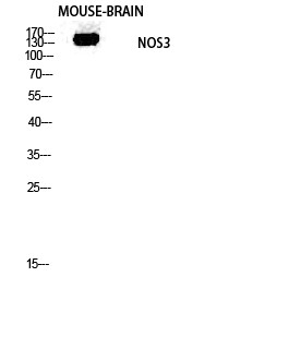
• Western blot analysis of mouse-brain lysis using NOS3 antibody. Antibody was diluted at 1:1000
  • Western blot analysis of A549 cells using NOS3 Polyclonal Antibody diluted at 1:1000
  • Western blot analysis of A549 cells using NOS3 Polyclonal Antibody diluted at 1:1000
  • Immunohistochemical analysis of paraffin-embedded Rat-spleen tissue. 1, NOS3 Polyclonal Antibody was diluted at 1:200 (4°C, overnight). 2, Sodium citrate pH 6.0 was used for antibody retrieval (>98°C, 20min). 3, Secondary antibody was diluted at 1:200 (room tempeRature, 30min). Negative control was used by secondary antibody only.
  • Immunohistochemical analysis of paraffin-embedded Mouse-testis tissue. 1, NOS3 Polyclonal Antibody was diluted at 1:200 (4°C, overnight). 2, Sodium citrate pH 6.0 was used for antibody retrieval (>98°C, 20min). 3, Secondary antibody was diluted at 1:200 (room tempeRature, 30min). Negative control was used by secondary antibody only.
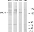
  • Western blot analysis of lysates from A549, K562, and COLO205 cells, using eNOS Antibody. The lane on the right is blocked with the synthesized peptide.
  • Immunofluorescence analysis of rat-lung tissue. 1, NOS3 Polyclonal Antibody (red) was diluted at 1:200 (4°C, overnight). 2, Cy3 labled Secondary antibody was diluted at 1:300 (room temperature, 50min).3, Picture B: DAPI (blue) 10min. Picture A:Target. Picture B: DAPI. Picture C: merge of A+B
  • Immunohistochemical analysis of paraffin-embedded Human-uterus-cancer tissue. 1, NOS3 Polyclonal Antibody was diluted at 1:200 (4°C, overnight). 2, Sodium citrate pH 6.0 was used for antibody retrieval (>98°C, 20min). 3, Secondary antibody was diluted at 1:200 (room tempeRature, 30min). Negative control was used by secondary antibody only.
  • Immunohistochemical analysis of paraffin-embedded Human-stomach tissue. 1, NOS3 Polyclonal Antibody was diluted at 1:200 (4°C, overnight). 2, Sodium citrate pH 6.0 was used for antibody retrieval (>98°C, 20min). 3, Secondary antibody was diluted at 1:200 (room tempeRature, 30min). Negative control was used by secondary antibody only.
  • Immunofluorescence analysis of rat-spleen tissue. 1, NOS3 Polyclonal Antibody (red) was diluted at 1:200 (4°C, overnight). 2, Cy3 labled Secondary antibody was diluted at 1:300 (room temperature, 50min).3, Picture B: DAPI (blue) 10min. Picture A:Target. Picture B: DAPI. Picture C: merge of A+B
  • Immunofluorescence analysis of rat-spleen tissue. 1, NOS3 Polyclonal Antibody (red) was diluted at 1:200 (4°C, overnight). 2, Cy3 labled Secondary antibody was diluted at 1:300 (room temperature, 50min).3, Picture B: DAPI (blue) 10min. Picture A:Target. Picture B: DAPI. Picture C: merge of A+B
  • Immunofluorescence analysis of rat-kidney tissue. 1, NOS3 Polyclonal Antibody (red) was diluted at 1:200 (4°C, overnight). 2, Cy3 labled Secondary antibody was diluted at 1:300 (room temperature, 50min).3, Picture B: DAPI (blue) 10min. Picture A:Target. Picture B: DAPI. Picture C: merge of A+B
  • Immunofluorescence analysis of rat-kidney tissue. 1, NOS3 Polyclonal Antibody (red) was diluted at 1:200 (4°C, overnight). 2, Cy3 labled Secondary antibody was diluted at 1:300 (room temperature, 50min).3, Picture B: DAPI (blue) 10min. Picture A:Target. Picture B: DAPI. Picture C: merge of A+B
  • Immunofluorescence analysis of rat-lung tissue. 1, NOS3 Polyclonal Antibody (red) was diluted at 1:200 (4°C, overnight). 2, Cy3 labled Secondary antibody was diluted at 1:300 (room temperature, 50min).3, Picture B: DAPI (blue) 10min. Picture A:Target. Picture B: DAPI. Picture C: merge of A+B
  • Western blot analysis of mouse-brain lysis using NOS3 antibody. Antibody was diluted at 1:1000
  • Immunohistochemical analysis of paraffin-embedded Rat-testis tissue. 1, NOS3 Polyclonal Antibody was diluted at 1:200 (4°C, overnight). 2, Sodium citrate pH 6.0 was used for antibody retrieval (>98°C, 20min). 3, Secondary antibody was diluted at 1:200 (room tempeRature, 30min). Negative control was used by secondary antibody only.
  • Immunofluorescence analysis of HeLa cells, using eNOS Antibody. The picture on the right is blocked with the synthesized peptide.
  • Immunohistochemical analysis of paraffin-embedded Rat-brain tissue. 1, NOS3 Polyclonal Antibody was diluted at 1:200 (4°C, overnight). 2, Sodium citrate pH 6.0 was used for antibody retrieval (>98°C, 20min). 3, Secondary antibody was diluted at 1:200 (room tempeRature, 30min). Negative control was used by secondary antibody only.
  • Western blot analysis of the lysates from HepG2 cells using eNOS antibody.
  • Immunofluorescence analysis of Hela cell. 1, NOS3 Polyclonal Antibody (green) was diluted at 1:200 (4°C overnight). 2, Goat Anti Rabbit Alexa Fluor 488 Catalog: (NA was diluted at 1:1000 (room temperature, 50min). 3 DAPI (blue) 10min.

Anti-NOS3 antibody (1145-1194) (STJ94539)

SKU:
STJ94539

Current Stock:
Host: Rabbit
Applications: WB/IHC/IF/ELISA
Reactivity: Human/Mouse/Rat
Note: STRICTLY FOR FURTHER SCIENTIFIC RESEARCH USE ONLY (RUO). MUST NOT TO BE USED IN DIAGNOSTIC OR THERAPEUTIC APPLICATIONS.
Short Description: Rabbit polyclonal antibody anti-Nitric oxide synthase 3 (1145-1194) is suitable for use in Western Blot, Immunohistochemistry, Immunofluorescence and ELISA research applications.
Clonality: Polyclonal
Conjugation: Unconjugated
Isotype: IgG
Formulation: Liquid in PBS containing 50% Glycerol, 0.5% BSA and 0.02% Sodium Azide.
Purification: The antibody was affinity-purified from rabbit antiserum by affinity-chromatography using epitope-specific immunogen.
Concentration: 1 mg/mL
Dilution Range: WB 1:500-2000
IF 1:50-300
IHC 1:50-300
Storage Instruction: Store at-20°C for up to 1 year from the date of receipt, and avoid repeat freeze-thaw cycles.
Gene Symbol: NOS3
Gene ID: 4846
Uniprot ID: NOS3_HUMAN
Immunogen Region: 1145-1194
Specificity: NOS3 Polyclonal Antibody detects endogenous levels of NOS3 protein.
Immunogen: The antiserum was produced against synthesized peptide derived from human eNOS. AA range:1145-1194
Tissue Specificity Platelets, placenta, liver and kidney.
Post Translational Modifications Phosphorylation by AMPK at Ser-1177 in the presence of Ca(2+)-calmodulin (CaM) activates activity. In absence of Ca(2+)-calmodulin, AMPK also phosphorylates Thr-495, resulting in inhibition of activity. Phosphorylation of Ser-114 by CDK5 reduces activity.
Function Produces nitric oxide (NO) which is implicated in vascular smooth muscle relaxation through a cGMP-mediated signal transduction pathway. NO mediates vascular endothelial growth factor (VEGF)-induced angiogenesis in coronary vessels and promotes blood clotting through the activation of platelets. Isoform eNOS13C: Lacks eNOS activity, dominant-negative form that may down-regulate eNOS activity by forming heterodimers with isoform 1.
Protein Name Nitric Oxide Synthase 3
Constitutive Nos
Cnos
Ec-Nos
Nos Type Iii
Nosiii
Nitric Oxide Synthase - Endothelial
Endothelial Nos
Enos
Database Links Reactome: R-HSA-1222556
Reactome: R-HSA-1474151
Reactome: R-HSA-203615
Reactome: R-HSA-203641
Reactome: R-HSA-203754
Reactome: R-HSA-392154
Reactome: R-HSA-5218920
Reactome: R-HSA-9009391
Cellular Localisation Cell Membrane
Membrane
Caveola
Cytoplasm
Cytoskeleton
Golgi Apparatus
Specifically Associates With Actin Cytoskeleton In The G2 Phase Of The Cell Cycle
Which Is Favored By Interaction With Nosip And Results In A Reduced Enzymatic Activity
Alternative Antibody Names Anti-Nitric Oxide Synthase 3 antibody
Anti-Constitutive Nos antibody
Anti-Cnos antibody
Anti-Ec-Nos antibody
Anti-Nos Type Iii antibody
Anti-Nosiii antibody
Anti-Nitric Oxide Synthase - Endothelial antibody
Anti-Endothelial Nos antibody
Anti-Enos antibody
Anti-NOS3 antibody

Information sourced from Uniprot.org

12 months for antibodies. 6 months for ELISA Kits. Please see website T&Cs for further guidance