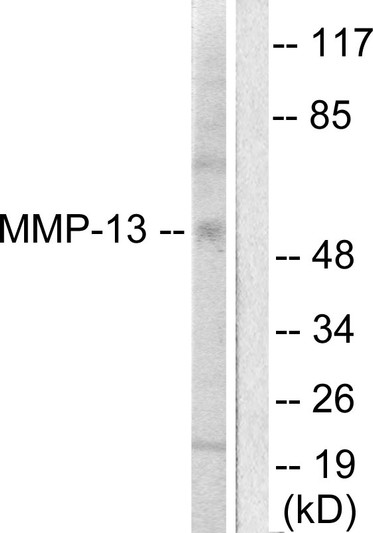
• Western blot analysis of lysates from LOVO cells, using MMP-13 Antibody. The lane on the right is blocked with the synthesized peptide.
  • Immunofluorescence analysis of HepG2 cells, using MMP-13 Antibody. The picture on the right is blocked with the synthesized peptide.
  • Immunohistochemistry analysis of paraffin-embedded human breast carcinoma tissue, using MMP-13 Antibody. The picture on the right is blocked with the synthesized peptide.
  • Immunohistochemical analysis of paraffin-embedded Human Mammary cancer. 1, Antibody was diluted at 1:200 (4°C overnight). 2, High-pressure and temperature EDTA, pH8.0 was used for antigen retrieval. 3, Secondary antibody was diluted at 1:200 (room temperature, 30min).
  • Immunohistochemical analysis of paraffin-embedded Human Mammary cancer. 1, Antibody was diluted at 1:200 (4°C overnight). 2, High-pressure and temperature EDTA, pH8.0 was used for antigen retrieval. 3, Secondary antibody was diluted at 1:200 (room temperature, 30min).
  • Immunohistochemical analysis of paraffin-embedded Human Colon cancer. 1, Antibody was diluted at 1:100 (4°C overnight). 2, High-pressure and temperature EDTA, pH8.0 was used for antigen retrieval. 3, Secondary antibody was diluted at 1:200 (room temperature, 30min).
  • Immunohistochemical analysis of paraffin-embedded Human Colon cancer. 1, Antibody was diluted at 1:100 (4°C overnight). 2, High-pressure and temperature EDTA, pH8.0 was used for antigen retrieval. 3, Secondary antibody was diluted at 1:200 (room temperature, 30min).
  • Western blot analysis of mouse-brain lysis using MMP-13 antibody. Antibody was diluted at 1:500
  • Western blot analysis of various cells using MMP-13 Polyclonal Antibody diluted at 1:500
  • Immunofluorescence analysis of Hela cell. 1, MMP-13 Polyclonal Antibody (green) was diluted at 1:200 (4°C overnight). 2, Goat Anti Rabbit Alexa Fluor 488 Catalog: (NA was diluted at 1:1000 (room temperature, 50min). 3 DAPI (blue) 10min.

Anti-MMP13 antibody (10-59) (STJ94161)

SKU:
STJ94161

Current Stock:
Host: Rabbit
Applications: WB/IHC/IF/ELISA
Reactivity: Human/Rat/Mouse
Note: STRICTLY FOR FURTHER SCIENTIFIC RESEARCH USE ONLY (RUO). MUST NOT TO BE USED IN DIAGNOSTIC OR THERAPEUTIC APPLICATIONS.
Short Description: Rabbit polyclonal antibody anti-Collagenase 3 (10-59) is suitable for use in Western Blot, Immunohistochemistry, Immunofluorescence and ELISA research applications.
Clonality: Polyclonal
Conjugation: Unconjugated
Isotype: IgG
Formulation: Liquid in PBS containing 50% Glycerol, 0.5% BSA and 0.02% Sodium Azide.
Purification: The antibody was affinity-purified from rabbit antiserum by affinity-chromatography using epitope-specific immunogen.
Concentration: 1 mg/mL
Dilution Range: WB 1:500-1:2000
IHC-p: 1:100-300
ELISA 1:20000
IF 1:100-300
Storage Instruction: Store at-20°C for up to 1 year from the date of receipt, and avoid repeat freeze-thaw cycles.
Gene Symbol: MMP13
Gene ID: 4322
Uniprot ID: MMP13_HUMAN
Immunogen Region: 10-59
Specificity: MMP-13 Polyclonal Antibody detects endogenous levels of MMP-13 protein.
Immunogen: The antiserum was produced against synthesized peptide derived from human MMP-13. AA range:10-59
Tissue Specificity Detected in fetal cartilage and calvaria, in chondrocytes of hypertrophic cartilage in vertebrae and in the dorsal end of ribs undergoing ossification, as well as in osteoblasts and periosteal cells below the inner periosteal region of ossified ribs. Detected in chondrocytes from in joint cartilage that have been treated with TNF and IL1B, but not in untreated chondrocytes. Detected in T lymphocytes. Detected in breast carcinoma tissue.
Post Translational Modifications The proenzyme is activated by removal of the propeptide.this cleavage can be effected by other matrix metalloproteinases, such as MMP2, MMP3 and MMP14 and may involve several cleavage steps. Cleavage can also be autocatalytic, after partial maturation by another protease or after treatment with 4-aminophenylmercuric acetate (APMA) (in vitro). N-glycosylated. Tyrosine phosphorylated by PKDCC/VLK.
Function Plays a role in the degradation of extracellular matrix proteins including fibrillar collagen, fibronectin, TNC and ACAN. Cleaves triple helical collagens, including type I, type II and type III collagen, but has the highest activity with soluble type II collagen. Can also degrade collagen type IV, type XIV and type X. May also function by activating or degrading key regulatory proteins, such as TGFB1 and CCN2. Plays a role in wound healing, tissue remodeling, cartilage degradation, bone development, bone mineralization and ossification. Required for normal embryonic bone development and ossification. Plays a role in the healing of bone fractures via endochondral ossification. Plays a role in wound healing, probably by a mechanism that involves proteolytic activation of TGFB1 and degradation of CCN2. Plays a role in keratinocyte migration during wound healing. May play a role in cell migration and in tumor cell invasion.
Protein Name Collagenase 3
Matrix Metalloproteinase-13
Mmp-13
Database Links Reactome: R-HSA-1442490
Reactome: R-HSA-1474228
Reactome: R-HSA-1592389
Reactome: R-HSA-2022090
Reactome: R-HSA-8941332
Cellular Localisation Secreted
Extracellular Space
Extracellular Matrix
Alternative Antibody Names Anti-Collagenase 3 antibody
Anti-Matrix Metalloproteinase-13 antibody
Anti-Mmp-13 antibody
Anti-MMP13 antibody

Information sourced from Uniprot.org

12 months for antibodies. 6 months for ELISA Kits. Please see website T&Cs for further guidance