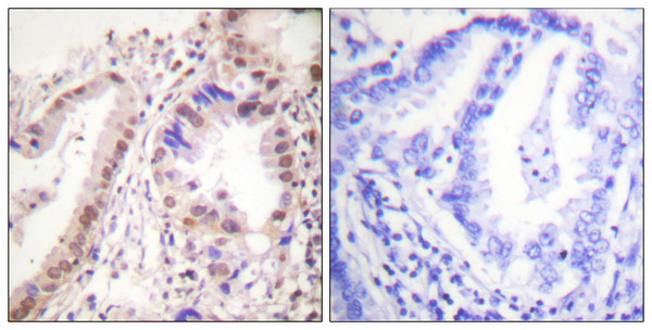
| Post Translational Modifications | Phosphorylated by cyclin A/CDK2 and CK1. Phosphorylation probably enhances transcriptional activity. Self-association induces phosphorylation. Dephosphorylation at Ser-118 by PPP5C inhibits its transactivation activity. Phosphorylated by LMTK3 in vitro. Glycosylated.contains N-acetylglucosamine, probably O-linked. Ubiquitinated.regulated by LATS1 via DCAF1 it leads to ESR1 proteasomal degradation. Deubiquitinated by OTUB1. Ubiquitinated by STUB1/CHIP.in the CA1 hippocampal region following loss of endogenous circulating estradiol (17-beta-estradiol/E2). Ubiquitinated by UBR5, leading to its degradation: UBR5 specifically recognizes and binds ligand-bound ESR1 when it is not associated with coactivators (NCOAs). In presence of NCOAs, the UBR5-degron is not accessible, preventing its ubiquitination and degradation. Dimethylated by PRMT1 at Arg-260. The methylation may favor cytoplasmic localization. Demethylated by JMJD6 at Arg-260. Palmitoylated (isoform 3). Not biotinylated (isoform 3). Palmitoylated by ZDHHC7 and ZDHHC21. Palmitoylation is required for plasma membrane targeting and for rapid intracellular signaling via ERK and AKT kinases and cAMP generation, but not for signaling mediated by the nuclear hormone receptor. |
| Function | Nuclear hormone receptor. The steroid hormones and their receptors are involved in the regulation of eukaryotic gene expression and affect cellular proliferation and differentiation in target tissues. Ligand-dependent nuclear transactivation involves either direct homodimer binding to a palindromic estrogen response element (ERE) sequence or association with other DNA-binding transcription factors, such as AP-1/c-Jun, c-Fos, ATF-2, Sp1 and Sp3, to mediate ERE-independent signaling. Ligand binding induces a conformational change allowing subsequent or combinatorial association with multiprotein coactivator complexes through LXXLL motifs of their respective components. Mutual transrepression occurs between the estrogen receptor (ER) and NF-kappa-B in a cell-type specific manner. Decreases NF-kappa-B DNA-binding activity and inhibits NF-kappa-B-mediated transcription from the IL6 promoter and displace RELA/p65 and associated coregulators from the promoter. Recruited to the NF-kappa-B response element of the CCL2 and IL8 promoters and can displace CREBBP. Present with NF-kappa-B components RELA/p65 and NFKB1/p50 on ERE sequences. Can also act synergistically with NF-kappa-B to activate transcription involving respective recruitment adjacent response elements.the function involves CREBBP. Can activate the transcriptional activity of TFF1. Also mediates membrane-initiated estrogen signaling involving various kinase cascades. Essential for MTA1-mediated transcriptional regulation of BRCA1 and BCAS3. Maintains neuronal survival in response to ischemic reperfusion injury when in the presence of circulating estradiol (17-beta-estradiol/E2). Isoform 3: Involved in activation of NOS3 and endothelial nitric oxide production. Isoforms lacking one or several functional domains are thought to modulate transcriptional activity by competitive ligand or DNA binding and/or heterodimerization with the full-length receptor. Binds to ERE and inhibits isoform 1. |
| Protein Name | Estrogen ReceptorErEr-AlphaEstradiol ReceptorNuclear Receptor Subfamily 3 Group A Member 1 |
| Database Links | Reactome: R-HSA-1251985Reactome: R-HSA-1257604Reactome: R-HSA-2219530Reactome: R-HSA-383280Reactome: R-HSA-4090294Reactome: R-HSA-5689896Reactome: R-HSA-6811558Reactome: R-HSA-8866910Reactome: R-HSA-8931987Reactome: R-HSA-8939211Reactome: R-HSA-8939256Reactome: R-HSA-8939902Reactome: R-HSA-9009391Reactome: R-HSA-9018519 |
| Cellular Localisation | Isoform 1: NucleusCytoplasmCell MembranePeripheral Membrane ProteinCytoplasmic SideA Minor Fraction Is Associated With The Inner MembraneIsoform 3: NucleusSingle-Pass Type I Membrane ProteinAssociated With The Inner Membrane Via Palmitoylation (Probable)At Least A Subset Exists As A Transmembrane Protein With A N-Terminal Extracellular DomainNucleusGolgi ApparatusColocalizes With Zdhhc7 And Zdhhc21 In The Golgi Apparatus Where Most Probably Palmitoylation OccursAssociated With The Plasma Membrane When Palmitoylated |
| Alternative Antibody Names | Anti-Estrogen Receptor antibodyAnti-Er antibodyAnti-Er-Alpha antibodyAnti-Estradiol Receptor antibodyAnti-Nuclear Receptor Subfamily 3 Group A Member 1 antibodyAnti-ESR1 antibodyAnti-ESR antibodyAnti-NR3A1 antibody |
Information sourced from Uniprot.org


