• Western blot analysis of extracts of various cell lines, using CAMKIV antibody (STJ11103655) at 1:1000 dilution. Secondary antibody: HRP Goat Anti-rabbit IgG (H+L) at 1:10000 dilution. Lysates/proteins: 25ug per lane. Blocking buffer: 3% non-fat dry milk in TBST. Detection: ECL Basic Kit. Exposure time: 30s.
  • Immunofluorescence analysis of U-2 OS cells using CAMKIV Rabbit monoclonal antibody (STJ11103655) at dilution of 1:100 (40x lens). Secondary antibody: Cy3 Goat Anti-Rabbit IgG (H+L) at 1:500 dilution. Blue: DAPI for nuclear staining.
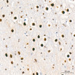
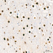
  • Immunohistochemistry of paraffin-embedded rat brain using CAMKIV rabbit monoclonal antibody (STJ11103655) at dilution of 1:100 (40x lens).
  • Immunofluorescence analysis of NIH-3T3 cells using CAMKIV Rabbit monoclonal antibody (STJ11103655) at dilution of 1:100 (40x lens). Secondary antibody: Cy3 Goat Anti-Rabbit IgG (H+L) at 1:500 dilution. Blue: DAPI for nuclear staining.
  • Immunohistochemistry of paraffin-embedded human esophageal cancer using CAMKIV rabbit monoclonal antibody (STJ11103655) at dilution of 1:100 (40x lens).
  • Immunohistochemistry analysis of CAMKIV in paraffin-embedded human breast cancer tissue using CAMKIV Rabbit monoclonal antibody (STJ11103655) at a dilution of 1:800 (40x lens). High pressure antigen retrieval was performed with 0. 01 M citrate buffer (pH 6. 0) prior to immunohistochemistry staining.
  • Immunohistochemistry of paraffin-embedded mouse spinal cord using CAMKIV rabbit monoclonal antibody (STJ11103655) at dilution of 1:100 (40x lens).
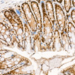
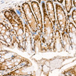
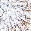
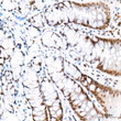
  • Immunohistochemistry analysis of CAMKIV in paraffin-embedded mouse colon tissue using CAMKIV Rabbit monoclonal antibody (STJ11103655) at a dilution of 1:800 (40x lens). High pressure antigen retrieval was performed with 0. 01 M citrate buffer (pH 6. 0) prior to immunohistochemistry staining.
  • Immunofluorescence analysis of NIH-3T3 cells using CAMKIV rabbit monoclonal antibody (STJ11103655) at dilution of 1:100 (40x lens). Blue: DAPI for nuclear staining.
  • Immunohistochemistry analysis of CAMKIV in paraffin-embedded rat brain tissue using CAMKIV Rabbit monoclonal antibody (STJ11103655) at a dilution of 1:800 (40x lens). High pressure antigen retrieval was performed with 0. 01 M citrate buffer (pH 6. 0) prior to immunohistochemistry staining.
  • Immunofluorescence analysis of U-2 OS cells using CAMKIV rabbit monoclonal antibody (STJ11103655) at dilution of 1:100 (40x lens). Blue: DAPI for nuclear staining.
  • Immunohistochemistry analysis of CAMKIV in paraffin-embedded rat colon tissue using CAMKIV Rabbit monoclonal antibody (STJ11103655) at a dilution of 1:800 (40x lens). High pressure antigen retrieval was performed with 0. 01 M citrate buffer (pH 6. 0) prior to immunohistochemistry staining.
  • Immunohistochemistry analysis of CAMKIV in paraffin-embedded mouse brain tissue using CAMKIV Rabbit monoclonal antibody (STJ11103655) at a dilution of 1:800 (40x lens). High pressure antigen retrieval was performed with 0. 01 M citrate buffer (pH 6. 0) prior to immunohistochemistry staining.
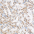
  • Immunohistochemistry analysis of CAMKIV in paraffin-embedded rat kidney tissue using CAMKIV Rabbit monoclonal antibody (STJ11103655) at a dilution of 1:800 (40x lens). High pressure antigen retrieval was performed with 0. 01 M citrate buffer (pH 6. 0) prior to immunohistochemistry staining.
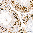
  • Immunohistochemistry analysis of CAMKIV in paraffin-embedded rat testis tissue using CAMKIV Rabbit monoclonal antibody (STJ11103655) at a dilution of 1:800 (40x lens). High pressure antigen retrieval was performed with 0. 01 M citrate buffer (pH 6. 0) prior to immunohistochemistry staining.
  • Western blot analysis of various lysates using CAMKIV Rabbit monoclonal antibody (STJ11103655) at 1:1000 dilution. Secondary antibody: HRP Goat Anti-Rabbit IgG (H+L) (STJS000856) at 1:10000 dilution. Lysates/proteins: 25 Mu g per lane. Blocking buffer: 3% nonfat dry milk in TBST. Detection: ECL Basic Kit. Exposure time: 30s.

Anti-CAMKIV antibody (374-473) [S5MR] (STJ11103655)

SKU:
STJ11103655

Shipping:
Free Shipping
Current Stock:
Host: Rabbit
Applications: WB/IHC/IF
Reactivity: Human/Mouse/Rat
Note: STRICTLY FOR FURTHER SCIENTIFIC RESEARCH USE ONLY (RUO). MUST NOT TO BE USED IN DIAGNOSTIC OR THERAPEUTIC APPLICATIONS.
Short Description: Rabbit monoclonal antibody anti-CAMKIV (374-473) is suitable for use in Western Blot, Immunohistochemistry and Immunofluorescence research applications.
Clonality: Monoclonal
Clone ID: S5MR
Conjugation: Unconjugated
Isotype: IgG
Formulation: PBS with 0.02% Sodium Azide, 0.05% BSA, 50% Glycerol, pH7.3.
Purification: Affinity purification
Dilution Range: WB 1:500-1:1000
IHC-P 1:50-1:200
IF/ICC 1:50-1:200
Storage Instruction: Store at-20°C for up to 1 year from the date of receipt, and avoid repeat freeze-thaw cycles.
Gene Symbol: CAMK4
Gene ID: 814
Uniprot ID: KCC4_HUMAN
Immunogen Region: 374-473
Immunogen: A synthetic peptide corresponding to a sequence within amino acids 374-473 of human CAMKIV (Q16566).
Immunogen Sequence: EGEKIQGDGAQAAVKGAQAE LMKVQALEKVKGADINAEEA PKMVPKAVEDGIKVADLELE EGLAEEKLKTVEEAAAPREG QGSSAVGFEVPQQDVILPEY
Tissue Specificity Expressed in brain, thymus, CD4 T-cells, testis and epithelial ovarian cancer tissue.
Post Translational Modifications Phosphorylated by CaMKK1 and CaMKK2 on Thr-200. Dephosphorylated by protein phosphatase 2A. Autophosphorylated on Ser-12 and Ser-13. Glycosylation at Ser-189 modulates the phosphorylation of CaMK4 at Thr-200 and negatively regulates its activity toward CREB1 in basal conditions and during early inomycin stimulation.
Function Calcium/calmodulin-dependent protein kinase that operates in the calcium-triggered CaMKK-CaMK4 signaling cascade and regulates, mainly by phosphorylation, the activity of several transcription activators, such as CREB1, MEF2D, JUN and RORA, which play pivotal roles in immune response, inflammation, and memory consolidation. In the thymus, regulates the CD4(+)/CD8(+) double positive thymocytes selection threshold during T-cell ontogeny. In CD4 memory T-cells, is required to link T-cell antigen receptor (TCR) signaling to the production of IL2, IFNG and IL4 (through the regulation of CREB and MEF2). Regulates the differentiation and survival phases of osteoclasts and dendritic cells (DCs). Mediates DCs survival by linking TLR4 and the regulation of temporal expression of BCL2. Phosphorylates the transcription activator CREB1 on 'Ser-133' in hippocampal neuron nuclei and contribute to memory consolidation and long term potentiation (LTP) in the hippocampus. Can activate the MAP kinases MAPK1/ERK2, MAPK8/JNK1 and MAPK14/p38 and stimulate transcription through the phosphorylation of ELK1 and ATF2. Can also phosphorylate in vitro CREBBP, PRM2, MEF2A and STMN1/OP18.
Protein Name Calcium/Calmodulin-Dependent Protein Kinase Type Iv
Camk Iv
Cam Kinase-Gr
Database Links Reactome: R-HSA-111932
Reactome: R-HSA-2151201
Reactome: R-HSA-442729
Reactome: R-HSA-9022535
Reactome: R-HSA-9022692
Reactome: R-HSA-9617324
Cellular Localisation Cytoplasm
Nucleus
Localized In Hippocampal Neuron Nuclei
In Spermatids
Associated With Chromatin And Nuclear Matrix
Alternative Antibody Names Anti-Calcium/Calmodulin-Dependent Protein Kinase Type Iv antibody
Anti-Camk Iv antibody
Anti-Cam Kinase-Gr antibody
Anti-CAMK4 antibody
Anti-CAMK antibody
Anti-CAMK-GR antibody
Anti-CAMKIV antibody

Information sourced from Uniprot.org

12 months for antibodies. 6 months for ELISA Kits. Please see website T&Cs for further guidance