| Host: |
Rabbit |
| Applications: |
WB/IHC/IF/ELISA |
| Reactivity: |
Human/Mouse/Rat |
| Note: |
STRICTLY FOR FURTHER SCIENTIFIC RESEARCH USE ONLY (RUO). MUST NOT TO BE USED IN DIAGNOSTIC OR THERAPEUTIC APPLICATIONS. |
| Short Description: |
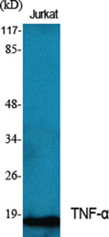
Rabbit polyclonal antibody anti-Tumor necrosis factor (141-190 aa) is suitable for use in Western Blot, Immunohistochemistry, Immunofluorescence and ELISA research applications. |
| Clonality: |
Polyclonal |
| Conjugation: |
Unconjugated |
| Isotype: |
IgG |
| Formulation: |
Liquid in PBS containing 50% Glycerol, 0.5% BSA and 0.02% Sodium Azide. |
| Purification: |
The antibody was affinity-purified from rabbit antiserum by affinity-chromatography using epitope-specific immunogen. |
| Concentration: |
1 mg/mL |
| Dilution Range: |
WB 1:500-1:2000IHC-P 1:100-300ELISA 1:20000IF 1:100-300 |
| Storage Instruction: |
Store at-20°C for up to 1 year from the date of receipt, and avoid repeat freeze-thaw cycles. |
| Gene Symbol: |
TNF |
| Gene ID: |
7124 |
| Uniprot ID: |
TNFA_HUMAN |
| Immunogen Region: |
141-190 aa |
| Specificity: |
TNF-Alpha Polyclonal Antibody detects endogenous levels of TNF-Alpha protein. |
| Immunogen: |
The antiserum was produced against synthesized peptide derived from the human TNFA at the amino acid range 141-190 |
| Function | Cytokine that binds to TNFRSF1A/TNFR1 and TNFRSF1B/TNFBR. It is mainly secreted by macrophages and can induce cell death of certain tumor cell lines. It is potent pyrogen causing fever by direct action or by stimulation of interleukin-1 secretion and is implicated in the induction of cachexia, Under certain conditions it can stimulate cell proliferation and induce cell differentiation. Impairs regulatory T-cells (Treg) function in individuals with rheumatoid arthritis via FOXP3 dephosphorylation. Up-regulates the expression of protein phosphatase 1 (PP1), which dephosphorylates the key 'Ser-418' residue of FOXP3, thereby inactivating FOXP3 and rendering Treg cells functionally defective. Key mediator of cell death in the anticancer action of BCG-stimulated neutrophils in combination with DIABLO/SMAC mimetic in the RT4v6 bladder cancer cell line. Induces insulin resistance in adipocytes via inhibition of insulin-induced IRS1 tyrosine phosphorylation and insulin-induced glucose uptake. Induces GKAP42 protein degradation in adipocytes which is partially responsible for TNF-induced insulin resistance. Plays a role in angiogenesis by inducing VEGF production synergistically with IL1B and IL6. Promotes osteoclastogenesis and therefore mediates bone resorption. The TNF intracellular domain (ICD) form induces IL12 production in dendritic cells. |
| Protein Name | Tumor Necrosis FactorCachectinTnf-AlphaTumor Necrosis Factor Ligand Superfamily Member 2Tnf-A Cleaved Into - Tumor Necrosis Factor - Membrane FormN-Terminal FragmentNtf - Intracellular Domain 1Icd1 - Intracellular Domain 2Icd2 - C-Domain 1 - C-Domain 2 - Tumor Necrosis Factor - Soluble Form |
| Database Links | Reactome: R-HSA-381340Reactome: R-HSA-5357786Reactome: R-HSA-5357905Reactome: R-HSA-5357956Reactome: R-HSA-5626978Reactome: R-HSA-5668541Reactome: R-HSA-6783783Reactome: R-HSA-6785807Reactome: R-HSA-75893 |
| Cellular Localisation | Cell MembraneSingle-Pass Type Ii Membrane ProteinTumor Necrosis FactorMembrane Form: MembraneSoluble Form: SecretedC-Domain 1: SecretedC-Domain 2: Secreted |
| Alternative Antibody Names | Anti-Tumor Necrosis Factor antibodyAnti-Cachectin antibodyAnti-Tnf-Alpha antibodyAnti-Tumor Necrosis Factor Ligand Superfamily Member 2 antibodyAnti-Tnf-A Cleaved Into - Tumor Necrosis Factor - Membrane Form antibodyAnti-N-Terminal Fragment antibodyAnti-Ntf - Intracellular Domain 1 antibodyAnti-Icd1 - Intracellular Domain 2 antibodyAnti-Icd2 - C-Domain 1 - C-Domain 2 - Tumor Necrosis Factor - Soluble Form antibodyAnti-TNF antibodyAnti-TNFA antibodyAnti-TNFSF2 antibody |
Information sourced from Uniprot.org
12 months for antibodies. 6 months for ELISA Kits. Please see website T&Cs for further guidance


