| Host: |
Rabbit |
| Applications: |
WB/IHC/IF |
| Reactivity: |
Human/Mouse/Rat |
| Note: |
STRICTLY FOR FURTHER SCIENTIFIC RESEARCH USE ONLY (RUO). MUST NOT TO BE USED IN DIAGNOSTIC OR THERAPEUTIC APPLICATIONS. |
| Short Description: |
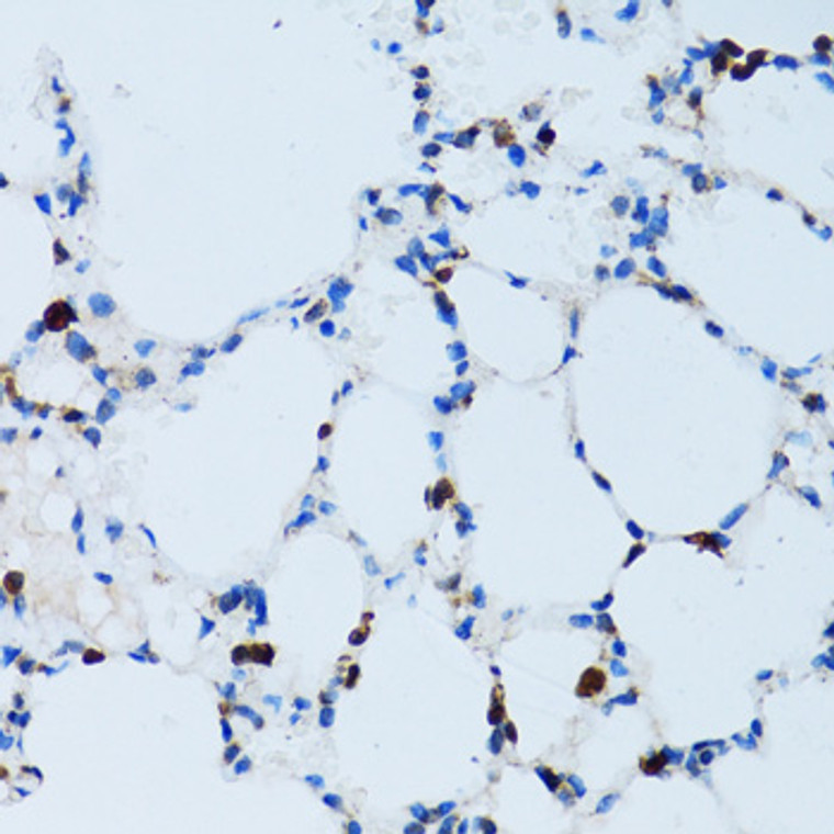
Rabbit polyclonal antibody anti-SPN (276-400) is suitable for use in Western Blot, Immunohistochemistry and Immunofluorescence research applications. |
| Clonality: |
Polyclonal |
| Conjugation: |
Unconjugated |
| Isotype: |
IgG |
| Formulation: |
PBS with 0.05% Proclin300, 50% Glycerol, pH7.3. |
| Purification: |
Affinity purification |
| Dilution Range: |
WB 1:100-1:500IHC-P 1:50-1:200IF/ICC 1:50-1:200 |
| Storage Instruction: |
Store at-20°C for up to 1 year from the date of receipt, and avoid repeat freeze-thaw cycles. |
| Gene Symbol: |
SPN |
| Gene ID: |
6693 |
| Uniprot ID: |
LEUK_HUMAN |
| Immunogen Region: |
276-400 |
| Immunogen: |
Recombinant fusion protein containing a sequence corresponding to amino acids 276-400 of human SPN (NP_003114.1). |
| Immunogen Sequence: |
LWRRRQKRRTGALVLSRGGK RNGVVDAWAGPAQVPEEGAV TVTVGGSGGDKGSGFPDGEG SSRRPTLTTFFGRRKSRQGS LAMEELKSGSGPSLKGEEEP LVASEDGAVDAPAPDEPEGG DGAAP |
| Tissue Specificity | Cell surface of thymocytes, T-lymphocytes, neutrophils, plasma cells and myelomas. |
| Post Translational Modifications | Glycosylated.has a high content of sialic acid and O-linked carbohydrate structures. Phosphorylation at Ser-355 is regulated by chemokines, requires its association with ERM proteins (EZR, RDX and MSN) and is essential for its function in the regulation of T-cell trafficking to lymph nodes. Has a high content of sialic acid and O-linked carbohydrate structures. Cleavage by CTSG releases its extracellular domain and triggers its intramembrane proteolysis by gamma-secretase releasing the CD43 cytoplasmic tail chain (CD43-ct) which translocates to the nucleus. CD43 cytoplasmic tail: Sumoylated. |
| Function | Predominant cell surface sialoprotein of leukocytes which regulates multiple T-cell functions, including T-cell activation, proliferation, differentiation, trafficking and migration. Positively regulates T-cell trafficking to lymph-nodes via its association with ERM proteins (EZR, RDX and MSN). Negatively regulates Th2 cell differentiation and predisposes the differentiation of T-cells towards a Th1 lineage commitment. Promotes the expression of IFN-gamma by T-cells during T-cell receptor (TCR) activation of naive cells and induces the expression of IFN-gamma by CD4(+) T-cells and to a lesser extent by CD8(+) T-cells. Plays a role in preparing T-cells for cytokine sensing and differentiation into effector cells by inducing the expression of cytokine receptors IFNGR and IL4R, promoting IFNGR and IL4R signaling and by mediating the clustering of IFNGR with TCR. Acts as a major E-selectin ligand responsible for Th17 cell rolling on activated vasculature and recruitment during inflammation. Mediates Th17 cells, but not Th1 cells, adhesion to E-selectin. Acts as a T-cell counter-receptor for SIGLEC1. CD43 cytoplasmic tail: Protects cells from apoptotic signals, promoting cell survival. |
| Protein Name | LeukosialinGpl115GalactoglycoproteinGalgpLeukocyte SialoglycoproteinSialophorinCd Antigen Cd43 Cleaved Into - Cd43 Cytoplasmic TailCd43-CtCd43ct |
| Database Links | Reactome: R-HSA-202733Reactome: R-HSA-210991 |
| Cellular Localisation | MembraneSingle-Pass Type I Membrane ProteinCell ProjectionMicrovillusUropodiumLocalizes To The Uropodium And Microvilli Via Its Interaction With Erm Proteins (EzrRdx And Msn)Cd43 Cytoplasmic Tail: NucleusNucleusPml BodyThe Sumoylated Form Localizes To The Pml Body |
| Alternative Antibody Names | Anti-Leukosialin antibodyAnti-Gpl115 antibodyAnti-Galactoglycoprotein antibodyAnti-Galgp antibodyAnti-Leukocyte Sialoglycoprotein antibodyAnti-Sialophorin antibodyAnti-Cd Antigen Cd43 Cleaved Into - Cd43 Cytoplasmic Tail antibodyAnti-Cd43-Ct antibodyAnti-Cd43ct antibodyAnti-SPN antibodyAnti-CD43 antibody |
Information sourced from Uniprot.org
12 months for antibodies. 6 months for ELISA Kits. Please see website T&Cs for further guidance


