• Western blot analysis of Hela (1), Rat brain (2), Rabbit Muscle (3), Sheep Muscle (4), and Mouse brain (5), diluted at 1:10000.
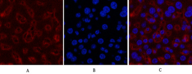
  • Immunofluorescence analysis of Human-colon tissue. 1, GAPDH monoclonal antibody (2B8) (red) was diluted at 1:200 (4°C, overnight). 2, Cy3 labled Secondary antibody was diluted at 1:300 (room temperature, 50min).3, Picture B: DAPI (blue) 10min. Picture A:Target. Picture B: DAPI. Picture C: merge of A+B
  • Immunofluorescence analysis of Mouse-liver tissue. 1, GAPDH monoclonal antibody (2B8) (red) was diluted at 1:200 (4°C, overnight). 2, Cy3 labled Secondary antibody was diluted at 1:300 (room temperature, 50min).3, Picture B: DAPI (blue) 10min. Picture A:Target. Picture B: DAPI. Picture C: merge of A+B
  • Immunohistochemical analysis of paraffin-embedded Mouse-heart tissue. 1, GAPDH monoclonal antibody (2B8) was diluted at 1:200 (4°C, overnight). 2, Sodium citrate pH 6.0 was used for antibody retrieval (>98°C, 20min). 3, Secondary antibody was diluted at 1:200 (room tempeRature, 30min). Negative control was used by secondary antibody only.
  • Immunohistochemical analysis of paraffin-embedded Rat-kidney tissue. 1, GAPDH monoclonal antibody (2B8) was diluted at 1:200 (4°C, overnight). 2, Sodium citrate pH 6.0 was used for antibody retrieval (>98°C, 20min). 3, Secondary antibody was diluted at 1:200 (room tempeRature, 30min). Negative control was used by secondary antibody only.
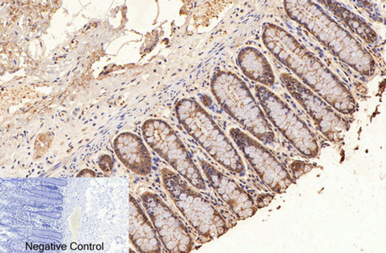
  • Immunohistochemical analysis of paraffin-embedded Human-colon tissue. 1, GAPDH monoclonal antibody (2B8) was diluted at 1:200 (4°C, overnight). 2, Sodium citrate pH 6.0 was used for antibody retrieval (>98°C, 20min). 3, Secondary antibody was diluted at 1:200 (room tempeRature, 30min). Negative control was used by secondary antibody only.
  • Western blot analysis of 1 HEK293 2 SW480 3 HEPG2 4 MCF-7 5 mouse brain 6 Rat brain 7 Hela 8 A549 lysates, primary antibody was diluted at 1:5000, 4°C over night, secondary antibody (cat: (NA was diluted at 1:10000, 37°C 1hour.
  • Immunofluorescence analysis of Hela cell. 1, Cyclin D1 Polyclonal Antibody (red) was diluted at 1:200 (4°C overnight). GAPDH monoclonal antibody (2B8) (green) was diluted at 1:200 (4°C overnight). 2, Goat Anti Rabbit Alexa Fluor 594 Catalog: (NA was diluted at 1:1000 (room temperature, 50min). Goat Anti Mouse Alexa Fluor 488 Catalog: (NA was diluted at 1:1000 (room temperature, 50min).

Anti-GAPDH antibody (200-300 aa) [2B8] (STJ96931)

SKU:
STJ96931

Current Stock:
Host: Mouse
Applications: WB/IF/IHC
Reactivity: Human/Rat/Mouse/Monkey/Dog/Chicken/Hamster/Rabbit/Pig/Sheep/Insect/Yeast/Cow
Note: STRICTLY FOR FURTHER SCIENTIFIC RESEARCH USE ONLY (RUO). MUST NOT TO BE USED IN DIAGNOSTIC OR THERAPEUTIC APPLICATIONS.
Short Description: Mouse monoclonal antibody anti-Glyceraldehyde-3-phosphate dehydrogenase (200-300 aa) is suitable for use in Western Blot, Immunofluorescence and Immunohistochemistry research applications.
Clonality: Monoclonal
Clone ID: 2B8
Conjugation: Unconjugated
Isotype: IgG1
Formulation: Liquid in PBS pH7.4, 0.5% BSA, 0.02% Sodium Azide and 50% Glycerol.
Purification: The antibody was affinity-purified from mouse ascites by affinity-chromatography using specific immunogen.
Dilution Range: WB 1:5000-20000
IHC 1:200-300
IF 1:200
Storage Instruction: Store at-20°C for up to 1 year from the date of receipt, and avoid repeat freeze-thaw cycles.
Gene Symbol: GAPDH
Gene ID: 2597
Uniprot ID: G3P_HUMAN
Immunogen Region: 200-300 aa
Specificity: The antibody detects endogenous GAPDH protein.
Immunogen: Synthetic Peptide of human GAPDH at the amino acid range 200-300
Function Has both glyceraldehyde-3-phosphate dehydrogenase and nitrosylase activities, thereby playing a role in glycolysis and nuclear functions, respectively. Glyceraldehyde-3-phosphate dehydrogenase is a key enzyme in glycolysis that catalyzes the first step of the pathway by converting D-glyceraldehyde 3-phosphate (G3P) into 3-phospho-D-glyceroyl phosphate. Modulates the organization and assembly of the cytoskeleton. Facilitates the CHP1-dependent microtubule and membrane associations through its ability to stimulate the binding of CHP1 to microtubules. Component of the GAIT (gamma interferon-activated inhibitor of translation) complex which mediates interferon-gamma-induced transcript-selective translation inhibition in inflammation processes. Upon interferon-gamma treatment assembles into the GAIT complex which binds to stem loop-containing GAIT elements in the 3'-UTR of diverse inflammatory mRNAs (such as ceruplasmin) and suppresses their translation. Also plays a role in innate immunity by promoting TNF-induced NF-kappa-B activation and type I interferon production, via interaction with TRAF2 and TRAF3, respectively. Participates in nuclear events including transcription, RNA transport, DNA replication and apoptosis. Nuclear functions are probably due to the nitrosylase activity that mediates cysteine S-nitrosylation of nuclear target proteins such as SIRT1, HDAC2 and PRKDC.
Protein Name Glyceraldehyde-3-Phosphate Dehydrogenase
Gapdh
Peptidyl-Cysteine S-Nitrosylase Gapdh
Database Links Reactome: R-HSA-70171
Reactome: R-HSA-70263
Cellular Localisation Cytoplasm
Cytosol
Nucleus
Perinuclear Region
Membrane
Cytoskeleton
Translocates To The Nucleus Following S-Nitrosylation And Interaction With Siah1
Which Contains A Nuclear Localization Signal
Postnuclear And Perinuclear Regions
Alternative Antibody Names Anti-Glyceraldehyde-3-Phosphate Dehydrogenase antibody
Anti-Gapdh antibody
Anti-Peptidyl-Cysteine S-Nitrosylase Gapdh antibody
Anti-GAPDH antibody
Anti-GAPD antibody
Anti-CDABP0047 antibody
Anti-OK antibody
Anti-SW-cl.12 antibody

Information sourced from Uniprot.org

12 months for antibodies. 6 months for ELISA Kits. Please see website T&Cs for further guidance