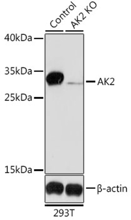
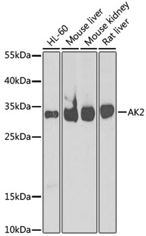
| Host: |
Rabbit |
| Applications: |
WB/IHC |
| Reactivity: |
Human/Mouse/Rat |
| Note: |
STRICTLY FOR FURTHER SCIENTIFIC RESEARCH USE ONLY (RUO). MUST NOT TO BE USED IN DIAGNOSTIC OR THERAPEUTIC APPLICATIONS. |
| Short Description: |
Rabbit polyclonal antibody anti-AK2 (1-232) is suitable for use in Western Blot and Immunohistochemistry research applications. |
| Clonality: |
Polyclonal |
| Conjugation: |
Unconjugated |
| Isotype: |
IgG |
| Formulation: |
PBS with 0.02% Sodium Azide, 50% Glycerol, pH7.3. |
| Purification: |
Affinity purification |
| Dilution Range: |
WB 1:500-1:2000IHC-P 1:50-1:200 |
| Storage Instruction: |
Store at-20°C for up to 1 year from the date of receipt, and avoid repeat freeze-thaw cycles. |
| Gene Symbol: |
AK2 |
| Gene ID: |
204 |
| Uniprot ID: |
KAD2_HUMAN |
| Immunogen Region: |
1-232 |
| Immunogen: |
Recombinant fusion protein containing a sequence corresponding to amino acids 1-232 of human AK2 (NP_001616.1). |
| Immunogen Sequence: |
MAPSVPAAEPEYPKGIRAVL LGPPGAGKGTQAPRLAENFC VCHLATGDMLRAMVASGSEL GKKLKATMDAGKLVSDEMVV ELIEKNLETPLCKNGFLLDG FPRTVRQAEMLDDLMEKRKE KLDSVIEFSIPDSLLIRRIT GRLIHPKSGRSYHEEFNPPK EPMKDDITGEPLIRRSDDNE KALKIRLQAYHTQTTPLIEY YRKRGIHSAIDASQTPDVVF ASILAAFSKATC |
| Tissue Specificity | Present in most tissues. Present at high level in heart, liver and kidney, and at low level in brain, skeletal muscle and skin. Present in thrombocytes but not in erythrocytes, which lack mitochondria. Present in all nucleated cell populations from blood, while AK1 is mostly absent. In spleen and lymph nodes, mononuclear cells lack AK1, whereas AK2 is readily detectable. These results indicate that leukocytes may be susceptible to defects caused by the lack of AK2, as they do not express AK1 in sufficient amounts to compensate for the AK2 functional deficits (at protein level). |
| Function | Catalyzes the reversible transfer of the terminal phosphate group between ATP and AMP. Plays an important role in cellular energy homeostasis and in adenine nucleotide metabolism. Adenylate kinase activity is critical for regulation of the phosphate utilization and the AMP de novo biosynthesis pathways. Plays a key role in hematopoiesis. |
| Protein Name | Adenylate Kinase 2 - MitochondrialAk 2Atp-Amp Transphosphorylase 2Atp -Amp PhosphotransferaseAdenylate Monophosphate Kinase Cleaved Into - Adenylate Kinase 2 - Mitochondrial - N-Terminally Processed |
| Database Links | Reactome: R-HSA-499943 |
| Cellular Localisation | Mitochondrion Intermembrane Space |
| Alternative Antibody Names | Anti-Adenylate Kinase 2 - Mitochondrial antibodyAnti-Ak 2 antibodyAnti-Atp-Amp Transphosphorylase 2 antibodyAnti-Atp -Amp Phosphotransferase antibodyAnti-Adenylate Monophosphate Kinase Cleaved Into - Adenylate Kinase 2 - Mitochondrial - N-Terminally Processed antibodyAnti-AK2 antibodyAnti-ADK2 antibody |
Information sourced from Uniprot.org
12 months for antibodies. 6 months for ELISA Kits. Please see website T&Cs for further guidance


