| Host: |
Rabbit |
| Applications: |
WB/IF |
| Reactivity: |
Human/Mouse/Rat |
| Note: |
STRICTLY FOR FURTHER SCIENTIFIC RESEARCH USE ONLY (RUO). MUST NOT TO BE USED IN DIAGNOSTIC OR THERAPEUTIC APPLICATIONS. |
| Short Description: |
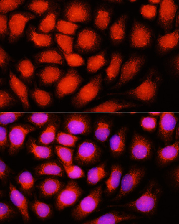
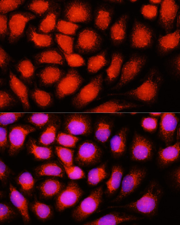
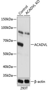
Rabbit polyclonal antibody anti-ACADVL (1-140) is suitable for use in Western Blot and Immunofluorescence research applications. |
| Clonality: |
Polyclonal |
| Conjugation: |
Unconjugated |
| Isotype: |
IgG |
| Formulation: |
PBS with 0.02% Sodium Azide, 50% Glycerol, pH7.3. |
| Purification: |
Affinity purification |
| Dilution Range: |
WB 1:500-1:2000IF/ICC 1:50-1:200 |
| Storage Instruction: |
Store at-20°C for up to 1 year from the date of receipt, and avoid repeat freeze-thaw cycles. |
| Gene Symbol: |
ACADVL |
| Gene ID: |
37 |
| Uniprot ID: |
ACADV_HUMAN |
| Immunogen Region: |
1-140 |
| Immunogen: |
Recombinant fusion protein containing a sequence corresponding to amino acids 1-140 of human ACADVL (NP_000009.1). |
| Immunogen Sequence: |
MQAARMAASLGRQLLRLGGG SSRLTALLGQPRPGPARRPY AGGAAQLALDKSDSHPSDAL TRKKPAKAESKSFAVGMFKG QLTTDQVFPYPSVLNEEQTQ FLKELVEPVSRFFEEVNDPA KNDALEMVEETTWQGLKELG |
| Tissue Specificity | Predominantly expressed in heart and skeletal muscle (at protein level). Also detected in kidney and liver (at protein level). |
| Post Translational Modifications | S-nitrosylation at Cys-237 in liver improves catalytic efficiency. |
| Function | Very long-chain specific acyl-CoA dehydrogenase is one of the acyl-CoA dehydrogenases that catalyze the first step of mitochondrial fatty acid beta-oxidation, an aerobic process breaking down fatty acids into acetyl-CoA and allowing the production of energy from fats. The first step of fatty acid beta-oxidation consists in the removal of one hydrogen from C-2 and C-3 of the straight-chain fatty acyl-CoA thioester, resulting in the formation of trans-2-enoyl-CoA. Among the different mitochondrial acyl-CoA dehydrogenases, very long-chain specific acyl-CoA dehydrogenase acts specifically on acyl-CoAs with saturated 12 to 24 carbons long primary chains. |
| Protein Name | Very Long-Chain Specific Acyl-Coa Dehydrogenase - MitochondrialVlcad |
| Database Links | Reactome: R-HSA-381038Reactome: R-HSA-77305 |
| Cellular Localisation | Mitochondrion Inner MembranePeripheral Membrane ProteinIsoform 2: Mitochondrion Inner Membrane |
| Alternative Antibody Names | Anti-Very Long-Chain Specific Acyl-Coa Dehydrogenase - Mitochondrial antibodyAnti-Vlcad antibodyAnti-ACADVL antibodyAnti-VLCAD antibody |
Information sourced from Uniprot.org
12 months for antibodies. 6 months for ELISA Kits. Please see website T&Cs for further guidance


