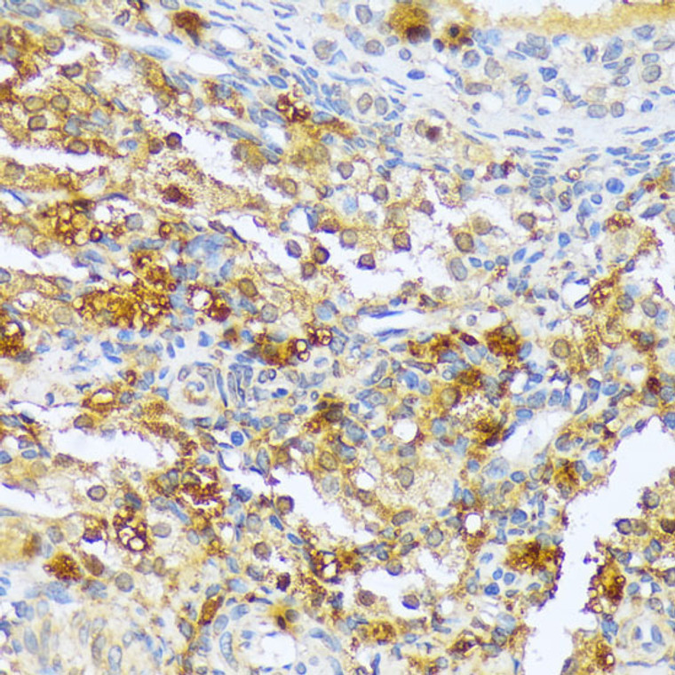
| Host: |
Rabbit |
| Applications: |
WB/IHC |
| Reactivity: |
Human/Mouse/Rat |
| Note: |
STRICTLY FOR FURTHER SCIENTIFIC RESEARCH USE ONLY (RUO). MUST NOT TO BE USED IN DIAGNOSTIC OR THERAPEUTIC APPLICATIONS. |
| Short Description: |
Rabbit polyclonal antibody anti-A1CF (50-120) is suitable for use in Western Blot and Immunohistochemistry research applications. |
| Clonality: |
Polyclonal |
| Conjugation: |
Unconjugated |
| Isotype: |
IgG |
| Formulation: |
PBS with 0.01% Thimerosal, 50% Glycerol, pH7.3. |
| Purification: |
Affinity purification |
| Dilution Range: |
WB 1:500-1:1000IHC-P 1:50-1:200 |
| Storage Instruction: |
Store at-20°C for up to 1 year from the date of receipt, and avoid repeat freeze-thaw cycles. |
| Gene Symbol: |
A1CF |
| Gene ID: |
29974 |
| Uniprot ID: |
A1CF_HUMAN |
| Immunogen Region: |
50-120 |
| Immunogen: |
Recombinant fusion protein containing a sequence corresponding to amino acids 50-120 of human A1CF (NP_055391.2). |
| Immunogen Sequence: |
APPERGCEIFIGKLPRDLFE DELIPLCEKIGKIYEMRMMM DFNGNNRGYAFVTFSNKVEA KNAIKQLNNYE |
| Tissue Specificity | Widely expressed with highest levels in brain, liver, pancreas, colon and spleen. |
| Function | Essential component of the apolipoprotein B mRNA editing enzyme complex which is responsible for the postranscriptional editing of a CAA codon for Gln to a UAA codon for stop in APOB mRNA. Binds to APOB mRNA and is probably responsible for docking the catalytic subunit, APOBEC1, to the mRNA to allow it to deaminate its target cytosine. The complex also protects the edited APOB mRNA from nonsense-mediated decay. |
| Protein Name | Apobec1 Complementation FactorApobec1-Stimulating Protein |
| Database Links | Reactome: R-HSA-72200Reactome: R-HSA-75094 |
| Cellular Localisation | NucleusEndoplasmic ReticulumCytoplasmPredominantly Nuclear Where It Localizes To HeterochromatinAlso Cytoplasmic Where It Is Found At The Outer Surface Of The Endoplasmic ReticulumShuttles Between The Nucleus And CytoplasmMay Be Transported Into The Nucleus By The Nuclear Import Protein Tnpo2/Trn2 Or By Apobec1 |
| Alternative Antibody Names | Anti-Apobec1 Complementation Factor antibodyAnti-Apobec1-Stimulating Protein antibodyAnti-A1CF antibodyAnti-ACF antibodyAnti-ASP antibody |
Information sourced from Uniprot.org
12 months for antibodies. 6 months for ELISA Kits. Please see website T&Cs for further guidance


